Primary Bone Lymphoma of the Distal Tibia Mimicking Brodie's Abscess
- Affiliations
-
- 1Department of Radiology, Hanyang University Seoul Hospital, Seoul, Korea. radsh@hanyang.ac.kr
- 2Department of, Pathology, Hanyang University Seoul Hospital, Seoul, Korea.
- KMID: 1897262
- DOI: http://doi.org/10.3348/jksr.2014.70.1.59
Abstract
- The "penumbra sign" on an unenhanced T1-weighted image is a well-known characteristic of Brodie's abscess, and this sign is extremely helpful for discriminating subacute osteomyelitis from other bone lesions. We present a case of primary bone lymphoma of the distal tibia mimicking subacute osteomyelitis with Brodie's abscess in a 50-year-old woman. Initial radiographs and MRI showed a lesion in the distal tibia consistent with Brodie's abscess with the penumbra sign. Histopathological examination of the surgical biopsy specimen confirmed the presence of a diffuse large B-cell lymphoma involving the bone.
MeSH Terms
Figure
-
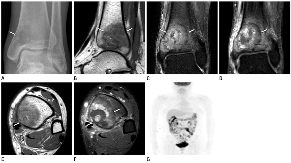
Fig. 1 A 50-year-old female with primary bone lymphoma. A. Plain radiograph of left ankle showing a ill-defined intramedullary osteolytic lesion in the distal tibia (arrow). B-F. Sagittal T1-weighted (B), coronal T2 SPAIR (C), coronal fat-saturated contrast-enhanced T1-weighted (D), axial T1-weighted (E) and axial fat-saturated contrast-enhanced T1-weighted (F) images of the distal tibia demonstrating the intramedullary mass with the penumbra sign (arrows). Note the hyperintense rim on T1-weighted image revealing the characteristic finding of the penumbra sign (arrowhead in B) and mild enhancement of the peri-osseous edema (arrowhead in F). G. MIP image from an PET/CT shows no evidence of other primary site or lymph node involvement. Note.-MIP = maximum intensity projection, PET/CT = positron emission tomography/CT, SPAIR = spectral attenuated inversion recovery

Fig. 2 Histology of the curettage specimen. A. The marrow space is replaced by diffuse sheet of large lymphoid cells with round to slightly indented, vesicular nuclei containing fine chromatin. The neoplastic lymphocytes have two to four nuclear membrane-bound nucleoli, pale eosinophilic to amphophilic cytoplasm. There are frequent mitotic figures and tingible body macrophages (H&E stain, × 400). B. Immunophenotypical analysis detected the expression of the B-correlated antigens; CD20 (× 400).
Reference
-
1. Afshar A, Mohammadi A. The "Penumbra Sign" on Magnetic Resonance Images of Brodie's Abscess: A Case Report. Iran J Radiol. 2011; 8:245–248.2. Schlur C, Bachy M, Wajfisz A, Ducou le Pointe H, Josset P, Vialle R. Osteoid osteoma mimicking Brodie's abscess in a 13-year-old girl. Pediatr Int. 2013; 55:e29–e31.3. Datir A, Lidder S, Pollock R, Tirabosco R, Saifuddin A. High-grade chondrosarcoma mimicking Brodie's abscess. Clin Radiol. 2009; 64:944–947.4. Bralić M, Stemberga V, Cuculić D, Coklo M, Bulić O, Grgurević E, et al. Primary non-Hodgkin's lymphoma of the humerus presenting as osteomyelitis. Coll Antropol. 2008; 32:Suppl 2. 229–231.5. Mika J, Schleicher I, Gerlach U, Adler CP, Uhl M, Knoeller SM. Primary bone lymphomas thought to be osteomyelitis urgently demand a rapid diagnosis in bone pathology. Anticancer Res. 2012; 32:4905–4912.6. Grey AC, Davies AM, Mangham DC, Grimer RJ, Ritchie DA. The 'penumbra sign' on T1-weighted MR imaging in subacute osteomyelitis: frequency, cause and significance. Clin Radiol. 1998; 53:587–592.7. Martí-Bonmatí L, Aparisi F, Poyatos C, Vilar J. Brodie abscess: MR imaging appearance in 10 patients. J Magn Reson Imaging. 1993; 3:543–546.8. Mulligan ME, McRae GA, Murphey MD. Imaging features of primary lymphoma of bone. AJR Am J Roentgenol. 1999; 173:1691–1697.9. Singh R, Al Wattar BH, Mohanty K. A rare case of primary bone lymphoma mimicking a pelvic abscess. Ann R Coll Surg Engl. 2011; 93:e141–e143.10. Krishnan A, Shirkhoda A, Tehranzadeh J, Armin AR, Irwin R, Les K. Primary bone lymphoma: radiographic-MR imaging correlation. Radiographics. 2003; 23:1371–1383. discussion 1384-1387.
