Imaging Findings of a Solitary Fibrous Tumor in Pancreas: A Case Report
- Affiliations
-
- 1Department of Radiology, Yeungnam University College of Medicine, Daegu, Korea. sungho1999@ynu.ac.kr
- KMID: 1897261
- DOI: http://doi.org/10.3348/jksr.2014.70.1.53
Abstract
- We report a case of a pathologically proven solitary fibrous tumor (SFT) of the pancreatic head. A 53-year-old woman was transferred our hospital for further evaluation of an incidental mass of the pancreatic head and computed tomography (CT) and magnetic resonance imaging (MRI) were performed. CT revealed that the mass had well defined margin with cystic and calcified portions on the pre-contrast scan and heterogeneously early strong and prolonged enhancement on contrast enhanced dynamic imaging with CT and MRI. Surgical resection was carried out. The mass was confirmed as a SFT arising from the pancreatic head, which is an extremely rare type of SFT. The imaging findings together with a brief literature review are described.
Figure
-
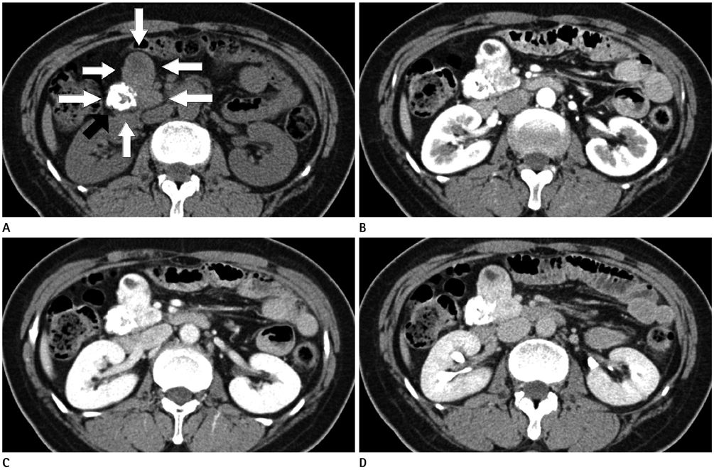
Fig. 1 CT findings of 53-year-old woman with solitary fibrous tumor in pancreatic head. A. An axial precontrast CT scan shows relatively well-defined, multilobulated mass (white arrows) in pancreatic head. The mass has calcified portion (black arrow). B-D. An axial contrast enhanced CT scans, the mass shows progressively heterogeneous enhancement during arterial (B) and portal phase (C). This mass still reveals slightly strong enhancement on delayed phase (D). Non-enhanced portions of mass indicate necrosis or cystic change.

Fig. 2 MRI findings of same patient. A-E. The MRI shows a low-signal-inensity lesion on the T1-weighted image (A), heterogeneously high signal intensity on the T2-weighted image (B), and heterogeneously low signal intensity on pre-contrast image (C), heterogeneously strong enhancement after contrast enhanced arterial (D) and veous phase (E) similar to that of CT scan.

Fig. 3 Histopathologic findings of specimen after surgical resection. A. Much cellular areas consist of spindle shaped cell with patternless cell deposition are seen at right left lower and right upper area. And central ossification area with low celluarity is also seen (H&E, × 100). B. In much celluar portion by higher magnification view, diffuse spindle shaped cell is seen (H&E, × 400). C. Specimen shows strong positive on immunohistochemical staining for CD34 (CD34, × 100).
Reference
-
1. Shanbhogue AK, Prasad SR, Takahashi N, Vikram R, Zaheer A, Sandrasegaran K. Somatic and visceral solitary fibrous tumors in the abdomen and pelvis: cross-sectional imaging spectrum. Radiographics. 2011; 31:393–408.2. Sugawara Y, Sakai S, Aono S, Takahashi T, Inoue T, Ohta K, et al. Solitary fibrous tumor of the pancreas. Jpn J Radiol. 2010; 28:479–482.3. Ginat DT, Bokhari A, Bhatt S, Dogra V. Imaging features of solitary fibrous tumors. AJR Am J Roentgenol. 2011; 196:487–495.4. Srinivasan VD, Wayne JD, Rao MS, Zynger DL. Solitary fibrous tumor of the pancreas: case report with cytologic and surgical pathology correlation and review of the literature. JOP. 2008; 9:526–530.5. Kwon HJ, Byun JH, Kang J, Park SH, Lee MG. Solitary fibrous tumor of the pancreas: imaging findings. Korean J Radiol. 2008; 9:Suppl. S48–S51.6. Daigeler A, Lehnhardt M, Langer S, Steinstraesser L, Steinau HU, Mentzel T, et al. Clinicopathological findings in a case series of extrathoracic solitary fibrous tumors of soft tissues. BMC Surg. 2006; 6:10.7. Tasdemir A, Soyuer I, Yurci A, Karahanli I, Akyildiz H. A huge solitary fibrous tumor localized in the pancreas: a young women. JOP. 2012; 13:304–307.8. Chetty R, Jain R, Serra S. Solitary fibrous tumor of the pancreas. Ann Diagn Pathol. 2009; 13:339–343.9. Miyamoto H, Molena DA, Schoeniger LO, Haodong Xu. Solitary fibrous tumor of the pancreas: a case report. Int J Surg Pathol. 2007; 15:311–314.10. Ishiwatari H, Hayashi T, Yoshida M, Kuroiwa G, Sato Y, Kobune M, et al. [A case of solitary fibrous tumor of the pancreas]. Nihon Shokakibyo Gakkai Zasshi. 2009; 106:1078–1085.
- Full Text Links
-
- Actions
-
Cited
- CITED
-
- Close
- Share
-
- Similar articles
-
- Solitary Fibrous Tumor of the Pancreas: Imaging Findings
- Solitary Fibrous Tumor of the Pancreas: A Case Report and Review of the Literature
- Solitary Fibrous Tumor of the Adrenal Gland: A Case Report
- Ultrasonographic Localization of Solitary Fibrous Tumor of Pleura: Case Report
- A Case of Solitary Fibrous Tumor in the Cheek
