Extremely rare case of extrahepatic duct phytobezoar treated with intraoperative transenteral endoscopy
- Affiliations
-
- 1Department of Surgery,Yeungnam University College of Medicine, Daegu, Korea. netetern@naver.com
- 2Department of Internal Medicine, Dongguk University Gyeongju Hospital, Gyeongju, Korea.
- KMID: 1882838
- DOI: http://doi.org/10.4174/astr.2014.87.2.100
Abstract
- Phytobezoar is a rare cause of gastro-intestinal tract obstruction. Common sites of phytobezoar are the stomach and small bowel. Naturally, extrahepatic duct phytobezoar is near impossible due to anatomical structure and location such as ampulla of vater, common bile duct and bifurcation of bile duct. Here, we present an extremely rare case of extrahepatic duct phytobezoar that resulted in abdominal pain. We successfully treated the case with extraoperative transenteral endoscopic removal of phytobezoar. For its great rarity and particular treatment approach, we report this case with review of literature.
Keyword
Figure
-
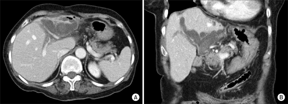
Fig. 1 The CT scan revealed dilatation and large stone of intrahepatic duct with air-biliary gram, parenchymal atrophy of left hepatic lobe and unremarkable right hepatic lobe. (A) Transverse section view. (B) Coronal section view.
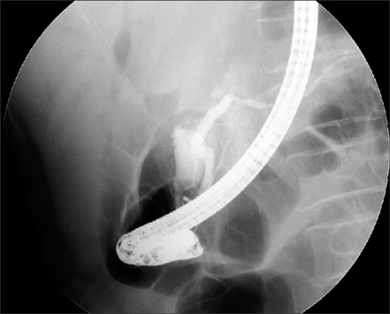
Fig. 2 Endoscopic retrograde cholangiopancreatography. There was not founded common bile duct.

Fig. 3 Schematic outline of operation field appearance.
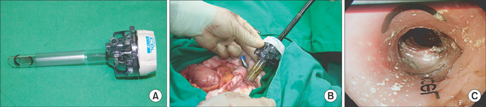
Fig. 4 Intraoperative transenteral endoscopy. (A) 15-mm Xcel trocar (Ethicon Inc., Somerville, NJ, USA). (B) Transenteral endoscopy using Trocar. (C) Inner appearance of transenteral endoscopy.

Fig. 5 Endoscopic procedure appearance. (A) There is a phytobezoar in hepatico-jejunostomy stoma. (B) Hepatico-jejunostomy stoma appearance after endoscopic removal of phytobezoar.

Fig. 6 The removed phytobezoar.
Reference
-
1. McKechnie JC. Gastroscopic removal of a phytobezoar. Gastroenterology. 1972; 62:1047–1051.2. Angelelli G, Magliocca M, Zaccheo N, Vinci R, Rotondo A. Intestinal obstruction caused by phytobezoar: computerized tomography findings: report of 3 cases. Radiol Med. 1997; 93:789–791.3. Robles R, Parrilla P, Escamilla C, Lujan JA, Torralba JA, Liron R, et al. Gastrointestinal bezoars. Br J Surg. 1994; 81:1000–1001.4. Singh SK, Marupaka SK. Duodenal date seed bezoar: a very unusual cause of partial gastric outlet obstruction. Australas Radiol. 2007; 51 Spec No.:B126–B129.5. Kim Y, Park BJ, Kim MJ, Sung DJ, Kim DS, Yu YD, et al. Biliary phytobezoar resulting in intestinal obstruction. World J Gastroenterol. 2013; 19:133–136.6. Lamotte M, Kockx M, Hautekeete M, Holvoet J, Hubens H. Biliary phytobezoar: a medical curiosity. Am J Gastroenterol. 1995; 90:1346–1348.7. Delabrousse E, Brunelle S, Saguet O, Destrumelle N, Landecy G, Kastler B. Small bowel obstruction secondary to phytobezoar CT findings. Clin Imaging. 2001; 25:44–46.8. Bae JM, Lee HK, Bae JD, Choi EA, Jung KH, Jung BW, et al. Trocar(R)(Ethicon) used intraoperative endoscopy in acute lower gastrointestinal bleeding. J Korean Surg Soc. 2004; 66:424–429.9. Ezzat RF, Rashid SA, Rashid AT, Abdullah KM, Ahmed SM. Small intestinal obstruction due to phytobezoar: a case report. J Med Case Rep. 2009; 3:9312.10. Ha SS, Lee HS, Jung MK, Jeon SW, Cho CM, Kim SK, et al. Acute intestinal obstruction caused by a persimmon phytobezoar after dissolution therapy with Coca-Cola. Korean J Intern Med. 2007; 22:300–303.
- Full Text Links
-
- Actions
-
Cited
- CITED
-
- Close
- Share
-
- Similar articles
-
- Extrahepatic Bile Duct Duplication with Intraductal Papillary Neoplasm: A Case Report
- Concurrent Yellow-to-white and Black Extrahepatic Bile Duct Stones
- A Rare Case of Extrahepatic Bile Duct Anomaly Associated with Multiple Stones
- A Case of Multiple Papillary Adenocarcinoma of the Extrahepatic Bile Duct : Findings of ERCP
- Biliary Web: A Rare Cause of Extrahepatic Biliary Obstruction
