An accessory limb with an imperforate anus
- Affiliations
-
- 1Department of Orthopaedic Surgery, Inje University Haeundae Paik Hospital, Busan, Korea.
- 2Department of Pathology, Inje University Haeundae Paik Hospital, Busan, Korea.
- 3Department of Pediatrics, Inje University Haeundae Paik Hospital, Busan, Korea.
- 4Department of Pediatric Surgery, Inje University Haeundae Paik Hospital, Busan, Korea. namsh@paik.ac.kr
- KMID: 1882828
- DOI: http://doi.org/10.4174/astr.2014.87.4.213
Abstract
- Congenital accessory limbs are very rare anomalies with many causative factors. We describe the case of a 1-day-old female neonate-born to a healthy, 27-year-old mother-who presented with an accessory limb (foot) attached to the buttock and an imperforate anus. We also provide a review of the relevant literature.
Keyword
Figure
-
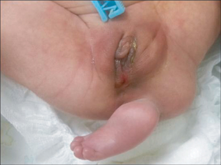
Fig. 1 A 1-day-old neonate presented with a congenital accessory limb attached to the buttock below an imperforate anus.

Fig. 2 An oval-shaped ossified bone was observed on the left side of the end of the sacrum.
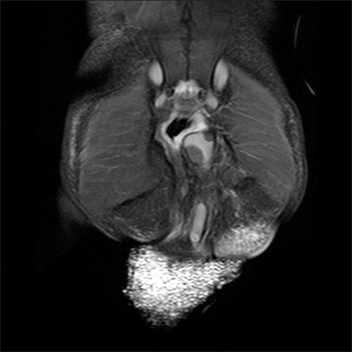
Fig. 3 A small round bone was located below the sacrum and a long bone was located below this small bone.
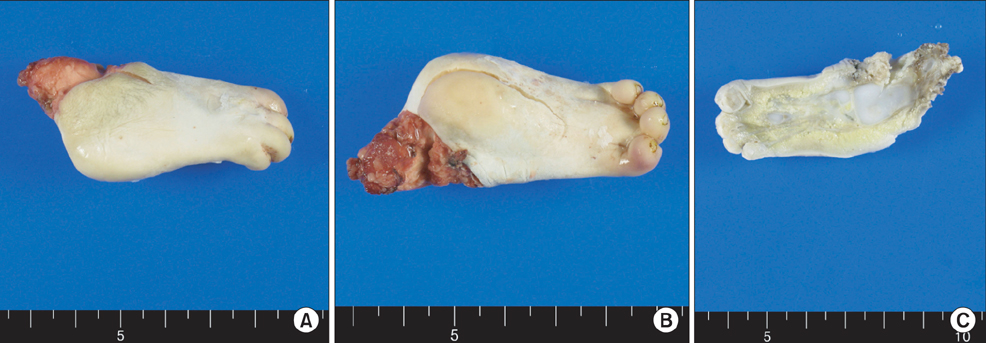
Fig. 4 Once excised, the accessory limb had a similar appearance to a normal neonatal foot, except for the fact that it only had 4 toes, with nails, which were bent towards the sole (A, B). The dissected surface indicated multiple bony structures that appeared similar to tarsal, metatarsal, cuneiform, talus, and malleolus bones. Skin and connective tissue were found to cover the bones (C).

Fig. 5 Under the microscope, the accessory foot was found to comprise ossified and nonossified bony structures, and the ossified bone had marrow space (A: H&E, ×20). Skeletal muscle bundles, vascular structures, and fat were found to cover the bones (B: H&E, ×40).

Fig. 6 An asymmetric buttock was noted on images obtained at the 8-month follow-up examination.
Cited by 1 articles
-
Polymelia (thoracomelia), an extremely rare appearance of congenital anomalic limb in a Nepalese child and its embryological basis
Ameet Kumar Jha, Samal Nauhria, Sabyasachi Maity
Anat Cell Biol. 2023;56(4):584-587. doi: 10.5115/acb.23.179.
Reference
-
1. O'Shea MK, Pillman SH, O'Shea R. Congenital third foot deformity: a case report. J Foot Ankle Surg. 2008; 47:583–588.2. Sharma L, Singh RB, Bhargava JS, Sharma VK. Accessory limbs with spinal lesions. Pediatr Surg Int. 1991; 6:227–229.3. Akyol D, Baltaci V, Kozinoglu H, Yuksel K, Kis S, Cicek N, et al. Accessory limb attached to the back. Turk J Med Sci. 1999; 29:199–201.4. Taniguchi K, Aoki Y, Kurimoto H, Okamura T. Baby with a third leg. J Pediatr Surg. 1975; 10:143–144.5. Chadha R, Bagga D, Malhotra CJ, Dhar A, Kumar A. Accessory limb attached to the back. J Pediatr Surg. 1993; 28:1615–1617.6. Rowe MI, Ravitch MM, Ranniger K. Operative correction of caudal duplication (dipygus). Surgery. 1968; 63:840–848.7. Unterscheider J, O'Byrne J, Foran A, Robinson I, Ryan S, Devaney D, et al. Prenatal identification of an accessory lower limb. Prenat Diagn. 2011; 31:1203–1204.8. Krishra A, Chandna S, Mishra NK, Gupta AK, Upadhyaya P. Accessory limb associated with spinal bifida. J Pediatr Surg. 1989; 24:604–606.9. Verma S, Khanna M, Tripathi VN, Yadav NC. Occurrence of polymelia in a female child. J Clin Imaging Sci. 2013; 3:18.
- Full Text Links
-
- Actions
-
Cited
- CITED
-
- Close
- Share
-
- Similar articles
-
- Two Experiences of Operation about Urogenital Anomalies Associated with Congenital Imperforate Anus
- A case of successful vaginal delivery in a patient with a repair of an imperforate anus
- Urogenital anomalies associated with imperforative anus
- A Case of Congenital Colonic Atresia Associated with Imperforate Anus
- Clinical analysis of imperforate anus
