Absence of transverse colon, persistent descending mesocolon, displaced small and large bowels: a rare congenital anomaly with a high risk of volvulus formation
- Affiliations
-
- 1Department of Anatomy, Melaka Manipal Medical College (Manipal Campus), Manipal University, Manipal, India. nayaksathish@gmail.com
- KMID: 1882609
- DOI: http://doi.org/10.5115/acb.2014.47.4.279
Abstract
- Congenital anomalies such as positional anomalies of the right half of the colon are more common when compared to its left half. We report a rare case of congenital anomaly where the transverse colon was totally absent. Ascending colon continued as descending colon at the right colic flexure. Ascending and descending colons formed an inverted U shaped loop which was situated in the right half of the abdomen. The sigmoid colon began from the descending colon, on the right side of the midline and coursed to the left iliac fossa. The terminal part of ascending colon and entire descending colon had a persistent mesocolon. The jejunum and ileum were situated in the upper left part of the abdominal cavity. This anomaly can cause volvulus of the colon at any stage of life. Furthermore, the knowledge of this anomaly is very useful for radiologists, gastroenterologists and surgeons.
Keyword
MeSH Terms
Figure
-
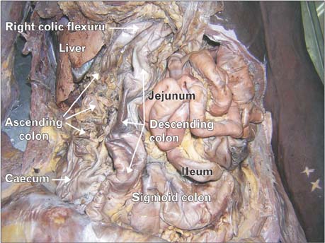
Fig. 1 Undisturbed position of abdominal viscera after reflection of anterior abdominal wall. Greater omentum has been reflected upwards. Note the inverted U shaped loop of ascending and descending colons. Jejunum and ileum are occupying the upper left part of the abdomen and the sigmoid colon is crossing from right to left.

Fig. 2 Descending colon (DC) has been pulled to the left to show the descending mesocolon (DMC). Note the continuity of the descending mesocolon with the ascending mesocolon (AMC), and sigmoid mesocolon (SMC). Ascending colon (AC) and sigmoid colon (SC) are also seen.
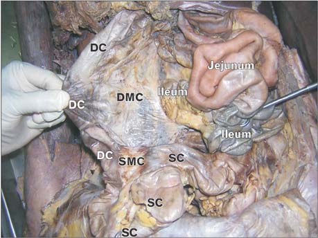
Fig. 3 The descending colon (DC) has been pulled to the right to show the descending mesocolon (DMC). Note the ileum passing behind the prominent margin of the descending mesocolon. Sigmoid colon (SC) and sigmoid mesocolon (SMC) are also seen.
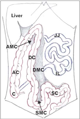
Fig. 4 A simplified illustration of the anomalies observed. The caecum (C), ascending colon (AC), ascending mesocolon (AMC), descending colon (DC), descending mesocolon (DMC), sigmoid colon (SC), sigmoid mesocolon (SMC), jejunum (JJ), and ileum (IL) have been labelled.
Reference
-
1. Vyas KC, Joshi CP, Misra S. Volvulus of descending colon with anomalous mesocolon. Indian J Gastroenterol. 1997; 16:34–35.2. Kedir M, Kotisso B, Messele G. Ileosigmoid knotting in Gondar teaching hospital north-west Ethiopia. Ethiop Med J. 1998; 36:255–260.3. Weledji EP, Theophile N. Primary small bowel volvulus in adults can be fatal: a report of two cases and brief review of the subject. Trop Doct. 2013; 43:75–76.4. Dutta AK. Essentials of human embryology. 8th ed. Calcutta: Current Books International;2008. p. 216.5. Papadimitriou G, Marinis A, Papakonstantinou A. Primary midgut volvulus in adults: report of two cases and review of the literature. J Gastrointest Surg. 2011; 15:1889–1892.6. Tarar JM, Ali MZ, Munir K. Descending colon volvulus: a case report and review of literature. J Sheikh Zayed Med Coll. 2011; 2:185–186.7. Shrivastava P, Tuli A, Kaur S, Raheja S. Right sided descending and sigmoid colon: its embryological basis and clinical implications. Anat Cell Biol. 2013; 46:299–302.8. Nayak SB, George BM, Mishra S, Surendran S, Shetty P, Shetty SD. Sessile ileum, subhepatic cecum, and uncinate appendix that might lead to a diagnostic dilemma. Anat Cell Biol. 2013; 46:296–298.
- Full Text Links
-
- Actions
-
Cited
- CITED
-
- Close
- Share
-
- Similar articles
-
- Transverse Mesocolic Hernia
- Anomalous peritoneal band connecting greater omentum to the ascending colon: a possible cause for dilation of ascending colon
- A Peterson's hernia and subsequent small bowel volvulus: surgical reconstruction utilizing transverse colon as a new Roux-en-Y limb - 1 case
- Right sided descending and sigmoid colon: its embryological basis and clinical implications
- Infected Colonic Duplication: A Case Report
