Extraskeletal Chondroma Causing Carpal Tunnel Syndrome: A Case Report
- Affiliations
-
- 1Department of Radiology, Haeundae Paik Hospital, Inje University College of Medicine, Busan, Korea. okkimmd@hanafos.com
- 2Department of Pathology, Haeundae Paik Hospital, Inje University College of Medicine, Busan, Korea.
- KMID: 1839436
- DOI: http://doi.org/10.3348/jksr.2014.70.3.217
Abstract
- Carpal tunnel syndrome caused by extraskeletal chondroma has been scarcely reported in the literature. Authors report a case of carpal tunnel syndrome as a result of an extraskeletal chondroma arising within the carpal tunnel, and describe the radiological and pathological findings of the mass. We also discuss the differential diagnosis of the calcified space, occupying lesions that may occur in carpal tunnel.
Figure
-
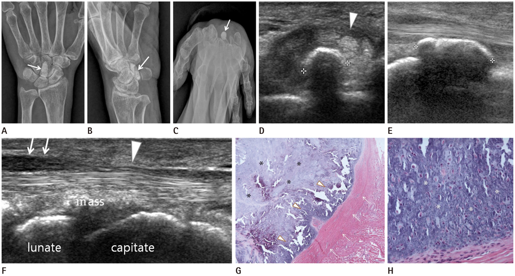
Fig. 1 A 64-year-old woman with carpal tunnel syndrome caused by an extraskeletal chondroma. A-C. AP (A) and oblique (B) radiographs of the wrist demonstrate a small, well-defined, oval, densely calcified mass (arrow) in the volar wrist. On carpal tunnel radiograph (C), the mass is located within the carpal tunnel (arrow). D-F. Transverse (D) and longitudinal (E) ultrasound (US) of the ventral wrist demonstrate a well-defined, echogenic lesion with posterior acoustic shadowing, consisting with a calcified lesion (cursors), in the deep portion of the carpal tunnel. Transverse (D) and paramedian longitudinal (F) US demonstrate that the median nerve flattens at the carpal tunnel (arrowhead in D, F) and appears to be hypoechoic swollen at the proximal portion (arrows in F). G, H. At low power photomicrograph (G), the tumor is encapsulated by a thick fibrous capsule and shows the nodular configuration (arrows). The hypocellular zone with myxoid background in the center (asterisks) and the hypercellular zone in the periphery (arrowheads) are seen (H&E stain, × 40). At high power photomicrograph (H), the peripheral zone of the tumor consists of mature benign looking chondrocytes with diffuse calcium depositions in the intercellular stroma (asterisks) (H&E stain, × 400).
Reference
-
1. Peetrons PA, Derbali W. Carpal tunnel syndrome. Semin Musculoskelet Radiol. 2013; 17:28–33.2. Kransdorf MJ, Meis JM. From the archives of the AFIP. Extraskeletal osseous and cartilaginous tumors of the extremities. Radiographics. 1993; 13:853–884.3. Chung EB, Enzinger FM. Chondroma of soft parts. Cancer. 1978; 41:1414–1424.4. Zlatkin MB, Lander PH, Begin LR, Hadjipavlou A. Soft-tissue chondromas. AJR Am J Roentgenol. 1985; 144:1263–1267.5. Cumming D, Massraf A, Jones JW. Extraskeletal chondroma as a cause of carpal tunnel syndrome: a case report. Hand Surg. 2005; 10:327–330.6. Bianchi S, Zwass A, Abdelwahab IF, Olivieri M, Marinaro E. Sonographic evaluation of soft tissue chondroma. J Clin Ultrasound. 1996; 24:148–150.7. Park JC, Lee YH, Jung KJ. Subungual hypervascular soft tissue chondroma mimicking a glomus tumor: a case report. J Korean Soc Ultrasound Med. 2009; 28:185–188.8. Bansal M, Goldman AB, DiCarlo EF, McCormack R. Soft tissue chondromas: diagnosis and differential diagnosis. Skeletal Radiol. 1993; 22:309–315.9. Fetsch JF, Vinh TN, Remotti F, Walker EA, Murphey MD, Sweet DE. Tenosynovial (extraarticular) chondromatosis: an analysis of 37 cases of an underrecognized clinicopathologic entity with a strong predilection for the hands and feet and a high local recurrence rate. Am J Surg Pathol. 2003; 27:1260–1268.10. Chen CK, Chung CB, Yeh L, Pan HB, Yang CF, Lai PH, et al. Carpal tunnel syndrome caused by tophaceous gout: CT and MR imaging features in 20 patients. AJR Am J Roentgenol. 2000; 175:655–659.
- Full Text Links
-
- Actions
-
Cited
- CITED
-
- Close
- Share
-
- Similar articles
-
- An Unusual Cause of Carpal Tunnel Syndrome: A Case of Tuberculosis of the Median Nerve
- Ultrasound-Guided Nerve Hydrodissection for Carpal Tunnel Syndrome
- Carpal Tunnel Syndrome in Children with Hypogammaglobulinemia: Case Report
- Thrombosed Persistent Median Artery with Bifid Median Nerve Causing Acute Carpal Tunnel Syndrome: A Case Report
- Extraskeletal Chondroma of Finger in a Young Woman: A Case Report
