Aberrant Right Vertebral Artery Originating from the Aortic Arch Distal to the Left Subclavian Artery: A Case Report
- Affiliations
-
- 1Department of Radiology, Haeundae Paik Hospital, Inje University College of Medicine, Busan, Korea. sartre81@gmail.com
- KMID: 1839430
- DOI: http://doi.org/10.3348/jksr.2014.70.3.191
Abstract
- We present a rare case of an aberrant right vertebral artery originated from the distal aortic arch. This issue has been incidentally detected on a preoperative CT angiography after a stabbing injury of the cervical spinal cord. Normally, the right vertebral artery originates from the right subclavian artery. Therefore, in this case report we will review the incidence and the embryological mechanism of this aberrant course of the right vertebral artery and we will discuss as well the clinical importance of this variation.
Figure
-
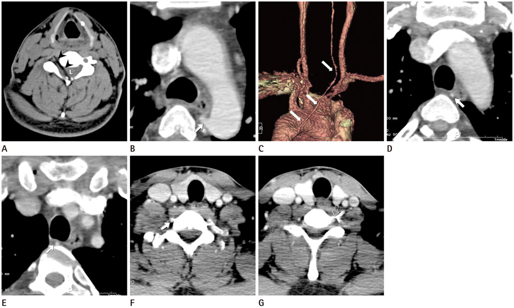
Fig. 1 A 34-year-old man with an incidentally diagnosed aberrant right vertebral artery. A. Unenhanced axial CT image reveals a linear, hypodense foreign body (chopstick, arrowheads) in the right central spinal canal (L) at C4-5 level caused by stabbing injury. B. Enhanced axial CT image reveals an aberrant origin of the right vertebral artery (RVA) with a Kummerell's diverticulum at its origin (arrow). C. Three dimensional volume rendering image demonstrates an anomalous origin of the RVA (arrows) that originating from the aortic arch distal to the left subclavian artery. D, E. Enhanced axial CT images shows an unusual course of the RVA. The prevertebral segment (arrows) is located in the retroesophageal and retrotracheal areas. F, G. Enhanced axial CT images reveal an aberrant entrance to the C7 transverse foramen of the RVA (arrow) which is compared with a normal entrance to the C6 transverse foramen of the left vertebral artery (open arrow).
Reference
-
1. Lemke AJ, Benndorf G, Liebig T, Felix R. Anomalous origin of the right vertebral artery: review of the literature and case report of right vertebral artery origin distal to the left subclavian artery. AJNR Am J Neuroradiol. 1999; 20:1318–1321.2. Nasir S, Hussain M, Khan SA, Mansoor MA, Sharif S. Anomalous origin of right vertebral artery from right external carotid artery. J Coll Physicians Surg Pak. 2010; 20:208–210.3. Albayram S, Gailloud P, Wasserman BA. Bilateral arch origin of the vertebral arteries. AJNR Am J Neuroradiol. 2002; 23:455–458.4. Ka-Tak W, Lam WW, Yu SC. MDCT of an aberrant right subclavian artery and of bilateral vertebral arteries with anomalous origins. AJR Am J Roentgenol. 2007; 188:W274–W275.5. Karcaaltincaba M, Haliloglu M, Ozkan E, Kocak M, Akinci D, Ariyurek M. Non-invasive imaging of aberrant right subclavian artery pathologies and aberrant right vertebral artery. Br J Radiol. 2009; 82:73–78.6. Karcaaltincaba M, Strottman J, Washington L. Multidetector-row CT angiographic findings in the bilateral aortic arch origin of the vertebral arteries. AJNR Am J Neuroradiol. 2003; 24:157.7. Newton TH, Mani RL. The vertebral artery. In : Newton TH, Potts DG, editors. Radiology of skull and Brain. St. Louis, MO: Mosby;1974. p. 1659–1672.8. Moore KL. The Developing Human: Clinically Oriented Embryology. 3rd ed. Philadelphia, PA: Saunders;1982.9. Goray VB, Joshi AR, Garg A, Merchant S, Yadav B, Maheshwari P. Aortic arch variation: a unique case with anomalous origin of both vertebral arteries as additional branches of the aortic arch distal to left subclavian artery. AJNR Am J Neuroradiol. 2005; 26:93–95.10. Dabus G, Walker MT. Right vertebral artery arising from the aortic arch distal to the left subclavian artery diagnosed with magnetic resonance angiography. Arch Neurol. 2010; 67:508.
- Full Text Links
-
- Actions
-
Cited
- CITED
-
- Close
- Share
-
- Similar articles
-
- Two Cases of Aberrant Vertebral Artery Originating from Aortic Arch Distal to Left Subclavian Artery
- Total Arch Replacement for Chronic Aortic Aneurysmal Dissection Patient with Aberrant Subclavian Artery
- Anomalous Origins of the Bilateral Vertebral Arteries Arising from the Aortic Arch: A Case Report
- Right aortic arch with aberrant left subclavian artery
- Surgical Treatment of Occluded Aberrant Left Subclavian Artery with Right-sided Aortic Arch: A case report
