CT Findings of Lymphoepithelioid Cell Lymphoma: A Case Report
- Affiliations
-
- 1Department of Radiology, Wonkwang University School of Medicine and Hospital, Iksan, Korea. yjyh@wonkwang.ac.kr
- 2Department of Radiology, Iksan Hospital, Iksan, Korea.
- 3Department of Pathology, Wonkwang University School of Medicine and Hospital, Iksan, Korea.
- KMID: 1839427
- DOI: http://doi.org/10.3348/jksr.2014.70.2.163
Abstract
- We report the computed tomography (CT) findings of a 70-year-old woman diagnosed with lymphoepithelioid lymphoma (Lennert's lymphoma). Neck, chest, and abdominal CT images revealed multiple enlarged lymph nodes, some of which showed heterogeneous mass or central low attenuation with peripheral rim enhancement. Although lymphoepithelioid cell lymphomas are very rare, they should be considered in the differential diagnosis of necrotic lymph nodes, particularly when combined with non-necrotic lymph nodes that show the typical radiologic features of lymphoma.
MeSH Terms
Figure
-
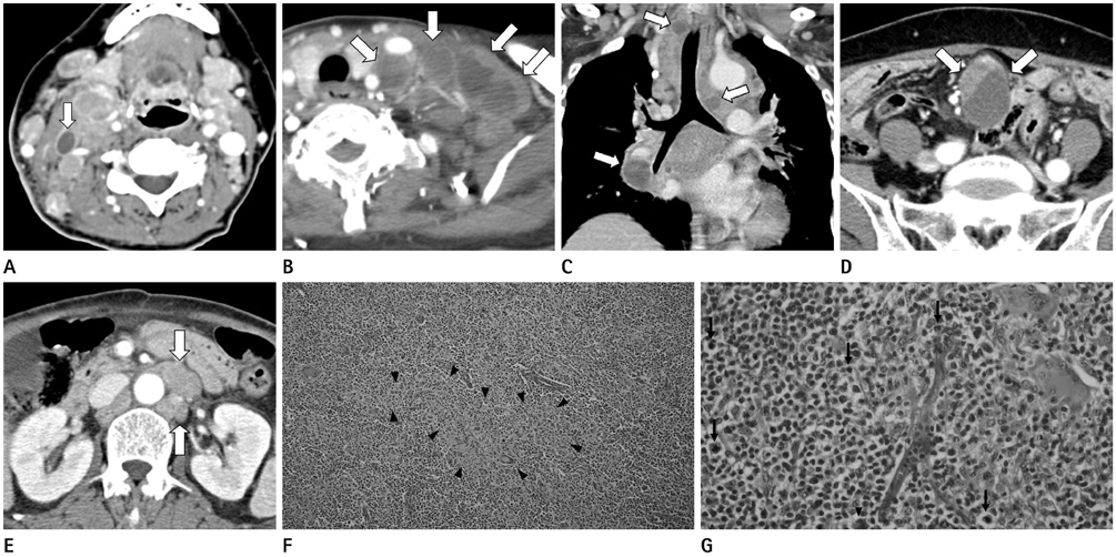
Fig. 1 Lymphoepithelioid cell lymphoma in a 70-year-old woman. A. Contrast-enhanced axial image of neck CT shows multiple enlarged lymph nodes in both neck spaces. The image demonstrates a necrotic lymph node at right level IIA (arrow). B. Contrast-enhanced axial image of chest CT shows multiple enlarged lymph nodes in both supraclavicular fossas. Note the central necroses in the left supraclavicular lymph nodes (arrows). C. Contrast-enhanced coronal image of chest CT also shows multiple necrotic lymph nodes with peripheral rim enhancement in the mediastinum and both pulmonary hila (arrows). D. Contrast-enhanced axial image of abdominal CT shows a markedly enlarged mesenteric lymph node (arrows) that contains solid and necrotic portions. E. Contrast-enhanced axial image of abdominal CT shows multiple enlarged lymph nodes (arrows). They show homogeneous enhancement and encase the mesenteric fat and vessels. F. Histopathologic exam shows neoplastic small cells that are admixed with confluent clusters of epithelioid histiocytes (arrowheads) (H&E, × 100). G. The tumor cells have nuclear irregularities and show frequent mitoses (arrows) and scattered Reed-Sternberg-like cells (arrowhead) (H&E, × 400).
Reference
-
1. Gödde-Salz E, Feller AC, Lennert K. Cytogenetic and immunohistochemical analysis of lymphoepithelioid cell lymphoma (Lennert's lymphoma): further substantiation of its T-cell nature. Leuk Res. 1986; 10:313–323.2. Jaffe ES, Harris NL, Stein H, Vardiman JW. World Health Organization Classification of Tumors. Pathology and genetics of tumors of haematopoietic and lymphoid tissues. Lyon: IARC Press;2001.3. Lennert K, Mestdagh J. [Hodgkin's disease with constantly high content of epithelioid cells]. Virchows Arch A Pathol Pathol Anat. 1968; 344:1–20.4. Daneshbod Y. Cytologic findings of peripheral T-cell lymphoma (PTCL) with high epitheloid cell content (Lennert's lymphoma) in imprint smear. A case report. Cytojournal. 2006; 3:3.5. Dong P, Wang B, Sun QY, Cui H. Tuberculosis versus non-Hodgkin's lymphomas involving small bowel mesentery: evaluation with contrast-enhanced computed tomography. World J Gastroenterol. 2008; 14:3914–3918.6. Yu RS, Zhang WM, Liu YQ. CT diagnosis of 52 patients with lymphoma in abdominal lymph nodes. World J Gastroenterol. 2006; 12:7869–7873.7. Choi JW, Kim SS, Kim EY, Heran M. Peripheral T-cell lymphoma in the neck: CT findings of lymph node involvement. AJNR Am J Neuroradiol. 2006; 27:1079–1082.8. Som PM. Lymph nodes of the neck. Radiology. 1987; 165:593–600.9. Hardy SM. The sandwich sign. Radiology. 2003; 226:651–652.10. Williams MP, Cherryman GR. Lymph-node calcification in Lennert's lymphoma. Br J Radiol. 1987; 60:1131–1132.
- Full Text Links
-
- Actions
-
Cited
- CITED
-
- Close
- Share
-
- Similar articles
-
- Fine Needle Aspiration Cytology of Peripheral T Cell Lymphoma, Lymphoepithelioid Cell Type: Report of A Case Mimicking Tuberculous Lymphadenitis
- Low Grade MALT Lymphoma of Rectum: A Case Report
- CT and (18F)FDG PET/CT Findings of Subcutaneous Panniculitis like T-Cell Lymphoma: A Case Report
- Spontaneous Perforation of Small Bowel Lymphoma Causing Massive Pneumoperitoneum: A case Report
- Sonographic Findings of Primary Tracheal Lymphoma: A Case Report
