Spontaneous Retropharyngeal Hematoma: A Case Report and Literature Overview
- Affiliations
-
- 1Department of Radiology, Haeundae Paik Hospital, Inje University College of Medicine, Busan, Korea. rjhrad@empal.com
- KMID: 1839417
- DOI: http://doi.org/10.3348/jksr.2014.70.2.87
Abstract
- A spontaneous retropharyngeal hematoma is a rare condition with a difficult diagnostic. This disease may rapidly progress to an airway obstruction. The author reports about a case of a 56-year-old man with an acute onset of sore throat, dysphonia and dyspnea. A retropharyngeal high attenuated soft tissue density could be seen on the neck CT. A rapid improvement of the retropharyngeal abnormality was seen on the 3 days follow-up MR imaging. Signal changes caused by blood products which were visible on the MRI images suggested the diagnosis of retropharyngeal hematoma. The patient was conservatively managed.
MeSH Terms
Figure
-
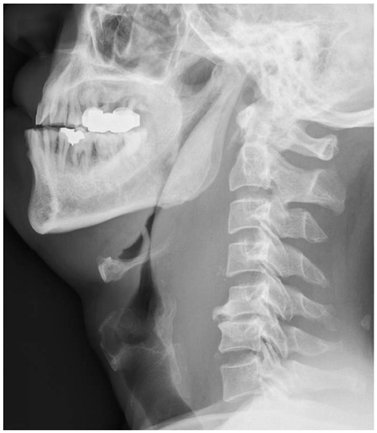
Fig. 1 Plain neck lateral view shows markedly increased thickness of retropharyngeal prevertebral soft tissue.
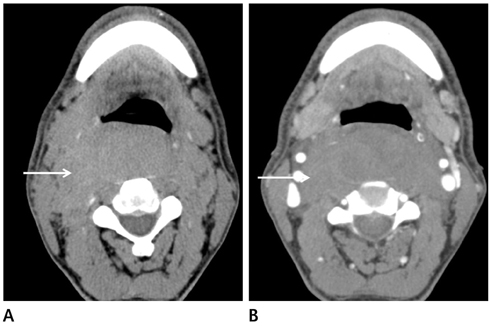
Fig. 2 Precontrast (A) neck computed tomography scan show an expansile mass lesion with slightly high attenuation as compared with muscle (arrow). Postcontrast CT (B) shows the high attenuated lesion (arrow) with slight peripheral enhancement in retropharyngeal space with narrowing of airway from occiput to C7 level.

Fig. 3 Magnetic resonance imaging. Axial spin-echo T1-weighted image (A) shows heterogenously high signal intensity lesion (arrow) in retropharyngeal space. Gadolinium enhanced T1-weighted image (B) show non-enhancing retropharyngeal collection (arrow). The airway is moderately compromised. On axial T2-weighted image (C), the lesion (arrow) shows low signal intensity, suggestive of blood products such as subacute hematoma. Sagittal T2-weighted image (D) shows low signal intensity of retropharyngeal hematoma (arrow).

Fig. 4 On 3-days follow-up check of plain neck lateral view, retropharyngeal prevertebral soft tissue markedly decreased in thickness.
Reference
-
1. Bloom DC, Haegen T, Keefe MA. Anticoagulation and spontaneous retropharyngeal hematoma. J Emerg Med. 2003; 24:389–394.2. Muñoz A, Fischbein NJ, de Vergas J, Crespo J, Alvarez-Vincent J. Spontaneous retropharyngeal hematoma: diagnosis by mr imaging. AJNR Am J Neuroradiol. 2001; 22:1209–1211.3. Kang SS, Jung SH, Kim MS, Hong SJ, Yoon YJ, Shin KM. Spontaneous retropharyngeal hematoma - a case report -. Korean J Pain. 2010; 23:211–214.4. Findlay JM, Belcher E, Black E, Sgromo B. Tracheo-oesophageal compression due to massive spontaneous retropharyngeal haematoma. Interact Cardiovasc Thorac Surg. 2013; 17:179–180.5. Singh A, Ofo E, Cumberworth V. Spontaneous retropharyngeal haematoma: a case report. J Med Case Rep. 2008; 2:8.6. al-Fallouji HK, Snow DG, Kuo MJ, Johnson PJ. Spontaneous retropharyngeal haematoma: two cases and a review of the literature. J Laryngol Otol. 1993; 107:649–650.7. Akoğlu E, Seyfeli E, Akoğlu S, Karazincir S, Okuyucu S, Dağli AS. Retropharyngeal hematoma as a complication of anticoagulation therapy. Ear Nose Throat J. 2008; 87:156–159.8. Keats TE, Sistrom C. Atlas of Radiologic Measurement. 7th ed. St. Louis, MO: Mosby;2001. p. 121.9. Rojas CA, Vermess D, Bertozzi JC, Whitlow J, Guidi C, Martinez CR. Normal thickness and appearance of the prevertebral soft tissues on multidetector CT. AJNR Am J Neuroradiol. 2009; 30:136–141.10. Som PM, Curtin HD. Head and neck imaging. 4th ed. St. Louis, MO: Mosby;2003. vol 2:p. 1505p. 1562–1564.
- Full Text Links
-
- Actions
-
Cited
- CITED
-
- Close
- Share
-
- Similar articles
-
- Spontaneous Retropharyngeal Hematoma: A Case Report
- Cervical Prevertebral Hematoma - a Rare Complication of Acupuncture Therapy: A Case Report
- Acute Airway Obstruction by a Retropharyngeal Hematoma when Performing Internal Jugular Vein Cannulation in a Patient with HELLP Syndrome: A case report
- Traumatic Retropharyngeal Hematoma of Delayed Onset in an Anticoagulated Patient
- A Clinical Review on Four Cases of the Retropharyngeal Hematoma
