Intracortical Chondroma: A Case Report
- Affiliations
-
- 1Department of Radiology, School of Medicine, Wonkwang University, Iksan, Korea. juhngsk@wonkwang.ac.kr
- 2Department of Pathology, School of Medicine, Wonkwang University, Iksan, Korea.
- KMID: 1823939
- DOI: http://doi.org/10.3348/jksr.2015.72.1.77
Abstract
- Herein we report a rare case of an intracortical chondroma with the histologic appearance of an enchondroma but located within the cortical bone.
MeSH Terms
Figure
-

Fig. 1 The whole body radioisotope bone scan shows a small focal hot uptake in the right distal femur (arrow).

Fig. 2 Anterior-posterior (A) and lateral (B) radiographs of the right knee show a focal lytic lesion with sclerotic rim and intralesional calcification in the right distal femur (arrows).

Fig. 3 The MRI shows an intracortical lesion with bright signal intensity on short tau inversion recovery (A) and high signal intensity and contrast enhancement of rim and internal focus on T2- (B) and contrast enhanced fat saturated T1-weighted (C) images in the right distal femur (arrows), suggestive of a cartilaginous lesion.
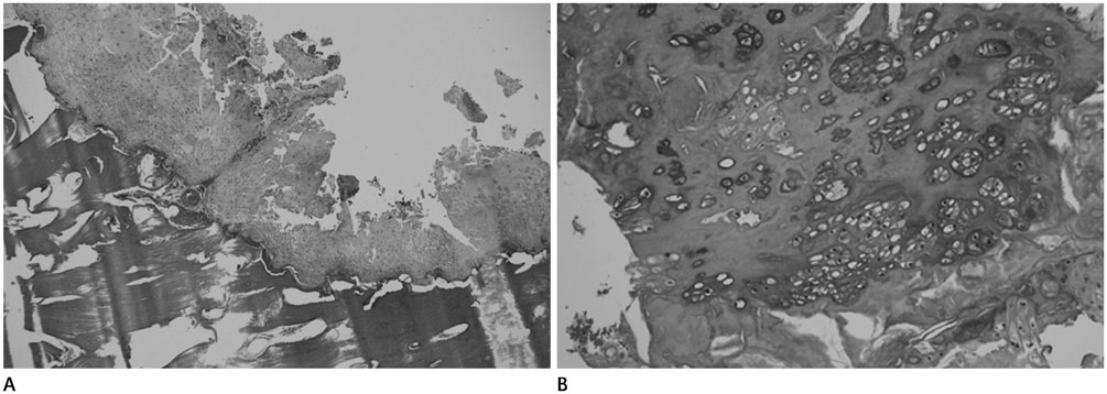
Fig. 4 The histopathology of the excised lesion from the right distal femur. A. Low magnification hematoxylin and eosin (H&E, × 40) staining demonstrates bland hyaline cartilage matrix and a benign interface with surrounding cortical bone. B. Higher magnification H&E staining (× 200) highlights the lone chondrocytes in the lacunae and the lack of nuclear atypia.
Reference
-
1. Abdelwahab IF, Hermann G, Lewis MM, Klein MJ. Case report 588: Intracortical chondroma of the left femur. Skeletal Radiol. 1990; 19:59–61.2. Rudman DP, Damron TA, Vermont A, Mathur S. Intracortical chondroma. Skeletal Radiol. 1998; 27:581–583.3. Ramnath RR, Rosenthal DI, Cates J, Gebhardt M, Quinn RH. Intracortical chondroma simulating osteoid osteoma treated by radiofrequency. Skeletal Radiol. 2002; 31:597–602.4. Jones KB, Buckwalter JA, Frassica FJ, McCarthy EF. Intracortical chondroma: a report of two cases. Skeletal Radiol. 2006; 35:298–301.5. Lui PC, Lau PP, Fan YS, Choi ST, Mak KH, Tam KF, et al. Intracortical chondroma in a 7-year-old boy and literature review. Pathology. 2006; 38:186–189.6. Choi E, Wert M, Guerrieri C, Tucci J. A pathologic fracture of an intracortical chondroma masking as an osteoid osteoma. Orthopedics. 2010; 33:845.7. Ruble C, Murphey MD, Fanburg-Smith J, Tyszko S, Zbojniewicz A, Franklin J. Imaging of intracortical chondroma. Special scientific paper session ISS 2009 Program Tuesday, September 1, 2009. Skeletal Radiol. 2009; 38:937–945.8. Ishida T, Goto T, Motoi N, Mukai K. Intracortical chondroblastoma mimicking intra-articular osteoid osteoma. Skeletal Radiol. 2002; 31:603–607.9. Hermann G, Klein MJ, Springfield D, Abdelwahab IF, Dan SJ. Intracortical osteosarcoma; two-year delay in diagnosis. Skeletal Radiol. 2002; 31:592–596.10. Fujiwara S, Nakamura I, Goto T, Motoi T, Yokokura S, Nakamura K. Intracortical chondromyxoid fibroma of humerus. Skeletal Radiol. 2003; 32:156–160.
- Full Text Links
-
- Actions
-
Cited
- CITED
-
- Close
- Share
-
- Similar articles
-
- Para-Falcine Chondroma: An Entity of Unacquaintance— A Case Report and Review of Literature
- Giant Extra-Capsular Synovial Chondroma of the knee joint: A Case Report
- Chondroma of Soft Tissue: A Case Report
- Chondromyxoid fibroma of the femur: a case report with intra-cortical location
- A Case of Auricular Chondroma
