Root coverage with a modified laterally positioned flap combined with a subepithelial connective tissue graft in advanced recession
- Affiliations
-
- 1Division of Periodontology, Department of Oral Medicine, Infection and Immunity, Harvard School of Dental Medicine, Boston, MA, USA.
- 2Graduate Institute of Clinical Dentistry, School of Dentistry, National Taiwan University, Taipei, Taiwan.
- 3Private Practice, Ottawa, ON, Canada.
- 4Division of Periodontics, Section of Oral and Diagnostic Sciences, College of Dental Medicine, Columbia University, New York, NY, USA. dr2267@columbia.edu
- KMID: 1820403
- DOI: http://doi.org/10.5051/jpis.2014.44.6.300
Abstract
- PURPOSE
A laterally positioned flap (LPF) combined with a subepithelial connective tissue graft (SCTG) is one of the conventional approaches for resolving gingival recession defects, with the advantages of flap flexibility and extended coverage of the tissue graft. However, thus far, evidence is lacking for the use of this technique for the treatment of advanced gingival recession defects. This report discusses three Miller class III cases with interproximal bone loss and wide and deep defects treated with a combination procedure of a modified laterally positioned flap (mLPF) and SCTG.
METHODS
mLPF combined with SCTG was performed for each case. The defect size and the degree of hypersensitivity at baseline and the final appointment in each case were documented.
RESULTS
The three cases had a mean initial defect of 7.7+/-1.5 mm and a mean residual defect of 1.7+/-1 mm at the 6-, 3-, and 36-month follow-up, respectively, after the root coverage surgery. The symptom of hypersensitivity was improved, and the patients were satisfied with the clinical outcomes.
CONCLUSIONS
The results demonstrated that the combination of mLPF with SCTG is promising for treating these advanced cases with respect to obtaining the expected root coverage with the gingival tissue.
Keyword
MeSH Terms
Figure
-
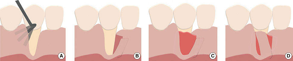
Figure 1 Surgical technique of modified laterally positioned flap combined with subepithelial connective tissue graft placement. (A) Use the instrument (e.g., Dr. Allen intrasulcular knife) to partially elevate the interproximal gingiva without opening the flap. The partially elevated area should be larger than the area where the tissue graft will be placed. (B) Incise and elevate the modified laterally positioned flap. The incision starts 2 mm below the zenith of the interproximal papilla and should cross the mucogingival junction in vertical and oblique directions to increase the flexibility. The flap is split in thickness, and the periosteum underneath the gingiva is intact. (C) Insert the subepithelial connective tissue graft underneath the partially and fully elevated gingiva. The upper border of the graft is positioned about 2-3 mm above the interproximal crestal bone level. (D) The flap is laterally positioned and sutured. The flap with sufficient flexibility should be positioned about 1-2 mm above the expected level of the future gingival margin.
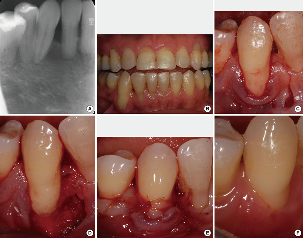
Figure 2 Case 1 clinical photographs. (A) The radiographic image of tooth #43; Tooth #43 had severe root resorption and significant interproximal bone loss. (B) The patient had generalized gingival recession and an open bite. Tooth #43 had a Miller class III gingival recession defect which was wide and deep (length: 8-9 mm). (C) A modified laterally positioned flap was made at the mesial interproximal gingiva with vertical and oblique incision. (D) The buccal alveolar bone of tooth #43 was missing and the resorption of the root apex was significant. (E) A clinical photograph on the date of completing surgery. (F) A clinical photograph at the six-month follow-up.

Figure 3 Case 2 clinical photographs. (A) Tooth #41 had a Miller class III gingival recession defect (length: 7-8 mm) and 0.5-1 mm wide buccal keratinized gingiva. (B) Tooth #41 had 1-2 mm of interproximal bone loss. (C) Modified laterally positioned flap was sutured and most of the subepithelial tissue graft was covered underneath the flap. (D) A clinical photograph at the three-month follow-up.

Figure 4 Case 3 clinical photograhs. (A) Periapical radiograph at the initial visit. (B) Bitewing radiograph at the initial visit. (C) Tooth #43 had a Miller class III gingival recession defect and the interproximal soft tissue had mild loss. (D) The flap was rotated mesially to assess the flexibility. (E) The subepithelial tissue graft was placed and sutured with Vicryl 5-0. (F) A clinical photograph on the date of completing surgery. (G) A clinical photograph at the six-month follow-up. (H) A clinical photograph of case 3 at the three-year follow-up.
Reference
-
1. Albandar JM, Kingman A. Gingival recession, gingival bleeding, and dental calculus in adults 30 years of age and older in the United States, 1988-1994. J Periodontol. 1999; 70:30–43.
Article2. Wennström JL. Mucogingival therapy. Ann Periodontol. 1996; 1:671–701.
Article3. Oates TW, Robinson M, Gunsolley JC. Surgical therapies for the treatment of gingival recession: a systematic review. Ann Periodontol. 2003; 8:303–320.
Article4. Cairo F, Nieri M, Pagliaro U. Efficacy of periodontal plastic surgery procedures in the treatment of localized facial gingival recessions: a systematic review. J Clin Periodontol. 2014; 41:Suppl 15. S44–S62.
Article5. Harvey PM. Management of advanced periodontitis. I. Preliminary report of a method of surgical reconstruction. N Z Dent J. 1965; 61:180–187.6. Grupe HE, Warren RF Jr. Repair of gingival defects by a sliding flap operation. J Periodontol. 1956; 27:92–95.
Article7. Nelson SW. The subpedicle connective tissue graft: a bilaminar reconstructive procedure for the coverage of denuded root surfaces. J Periodontol. 1987; 58:95–102.8. Borghetti A, Louise F. Controlled clinical evaluation of the subpedicle connective tissue graft for the coverage of gingival recession. J Periodontol. 1994; 65:1107–1112.
Article9. Smukler H. Laterally positioned mucoperiosteal pedicle grafts in the treatment of denuded roots: a clinical and statistical study. J Periodontol. 1976; 47:590–595.
Article10. Caffesse RG, Guinard EA. Treatment of localized gingival recessions. Part IV. Results after three years. J Periodontol. 1980; 51:167–170.
Article11. Caffesse RG, Alspach SR, Morrison EC, Burgett FG. Lateral sliding flaps with and without citric acid. Int J Periodontics Restorative Dent. 1987; 7:42–57.12. Zucchelli G, Cesari C, Amore C, Montebugnoli L, De Sanctis M. Laterally moved, coronally advanced flap: a modified surgical approach for isolated recession-type defects. J Periodontol. 2004; 75:1734–1741.
Article13. Santana RB, Furtado MB, Mattos CM, de Mello Fonseca E, Dibart S. Clinical evaluation of single-stage advanced versus rotated flaps in the treatment of gingival recessions. J Periodontol. 2010; 81:485–492.
Article14. Miller PD Jr. A classification of marginal tissue recession. Int J Periodontics Restorative Dent. 1985; 5:8–13.15. Cairo F, Cortellini P, Tonetti M, Nieri M, Mervelt J, Cincinelli S, et al. Coronally advanced flap with and without connective tissue graft for the treatment of single maxillary gingival recession with loss of inter-dental attachment. A randomized controlled clinical trial. J Clin Periodontol. 2012; 39:760–768.
Article16. Miller SC. Textbook of periodontia. Philadelphia: Blakiston Co.;1938.17. Allen AL. Use of the supraperiosteal envelope in soft tissue grafting for root coverage. II. Clinical results. Int J Periodontics Restorative Dent. 1994; 14:302–315.18. Ricci G, Silvestri M, Rasperini G, Cattaneo V. Root coverage: a clinical/statistical comparison between subpedicle connective tissue graft and laterally positioned full thickness flaps. J Esthet Dent. 1996; 8:66–73.
Article19. Han JS, John V, Blanchard SB, Kowolik MJ, Eckert GJ. Changes in gingival dimensions following connective tissue grafts for root coverage: comparison of two procedures. J Periodontol. 2008; 79:1346–1354.
Article20. Bouchard P, Malet J, Borghetti A. Decision-making in aesthetics: root coverage revisited. Periodontol 2000. 2001; 27:97–120.
Article21. Langer B, Langer L. Subepithelial connective tissue graft technique for root coverage. J Periodontol. 1985; 56:715–720.
Article22. Gordon HP, Sullivan HC, Atkins JH. Free autogenous gingival grafts. II. Supplemental findings: histology of the graft site. Periodontics. 1968; 6:130–133.23. Esteibar JR, Zorzano LA, Cundin EE, Blanco JD, Medina JR. Complete root coverage of Miller Class III recessions. Int J Periodontics Restorative Dent. 2011; 31:e1–e7.24. Chambrone LA, Chambrone L. Treatment of Miller class I and II localized recession defects using laterally positioned flaps: a 24-month study. Am J Dent. 2009; 22:339–344.25. Zucchelli G, Marzadori M, Mele M, Stefanini M, Montebugnoli L. Root coverage in molar teeth: a comparative controlled randomized clinical trial. J Clin Periodontol. 2012; 39:1082–1088.
Article26. Yilmaz E, Ozcelik O, Comert M, Ozturan S, Seydaoglu G, Teughels W, et al. Laser-assisted laterally positioned flap operation: a randomized controlled clinical trial. Photomed Laser Surg. 2014; 32:67–74.
Article
- Full Text Links
-
- Actions
-
Cited
- CITED
-
- Close
- Share
-
- Similar articles
-
- Laterally positioned flap using subepithelial connective tissue graft for iatrogenic gingival recession treatment
- Root coverage using laterally positioned flap and subepithelial connective tissue graft for the treatment of the isolated recession defects on mandibular anterior teeth: case report
- Root coverage with subeptithelial connective tissue grafts
- The evaluation of clinical outcomes on various procedures using subepithelial connective tissue graft for coverage of gingival recession
- Root coverage using subepithelial connective tissue graft
