Adrenal ganglioneuroma with hepatic metastasis
- Affiliations
-
- 1Department of Pathology, Keimyung University School of Medicine, Daegu, Korea. yunakang@dsmc.or.kr
- 2Department of Surgery, Keimyung University School of Medicine, Daegu, Korea.
- 3Department of Radiology, Keimyung University School of Medicine, Daegu, Korea.
- KMID: 1820022
- DOI: http://doi.org/10.4174/jkss.2011.80.4.297
Abstract
- Ganglioneuroma is the most differentiated tumor of neural crest origin and rarely arises in the adrenal gland. Ganglioneuroma is typically known to be benign, but very rarely can metastasize to distant sites. We report a case of a 31-year-old man with a huge adrenal mass with hepatic metastases.
Keyword
Figure
-

Fig. 1 Axial magnetic resonance images show a large mass in the right adrenal gland, which reveals hypointense signal intensity on T1-weighted image (A) and slightly hyperintense and heterogeneous signal intensity on T2-weighted image (B).

Fig. 2 Axial T1-weighted axial magnetic resonace image shows a small wedge-shaped hypointense nodule in the subcapsular location of medial segment (arrow).
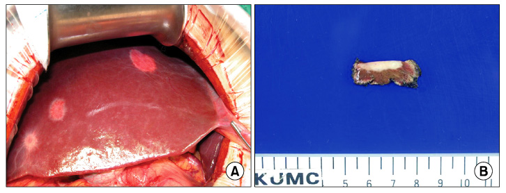
Fig. 3 The liver shows three white and round subcapsular metastatic lesions (A). Cut surface of one of the wedge resected liver tissue shows pale tan to white scar-like lesion with infiltrative border at subcapsular area (B).

Fig. 4 Cut surface of the right adrenal gland showed a round well demarcated pale tan, solid, and firm mass.

Fig. 5 Section of the adrenal mass shows scattered ganglion cells surrounded by fascicles of schwann-like spindle cells (A, H&E, ×100). There is a focus of differentiating neuroblasts (B, H&E, ×200).

Fig. 6 Section of the liver tissue shows scar-like lesion at low magnification (A, H&E, ×40) and scattered ganglion cells surrounded by fascicles of schwann-like spindle cells at high magnification (B, H&E, ×100).
Reference
-
1. Banks E, Yum M, Brodhecker C, Goheen M. A malignant peripheral nerve sheath tumor in association with a paratesticular ganglioneuroma. Cancer. 1989. 64:1738–1742.2. Ambros IM, Zellner A, Roald B, Amann G, Ladenstein R, Printz D, et al. Role of ploidy, chromosome 1p, and Schwann cells in the maturation of neuroblastoma. N Engl J Med. 1996. 334:1505–1511.3. Srinivasan R, Koliyadan KS, Krishnand G, Bhat SS. Retroperitoneal ganglioneuroma with lymphnode metastasis: a case report. Indian J Pathol Microbiol. 2007. 50:32–35.4. Joshi VV. Peripheral neuroblastic tumors: pathologic classification based on recommendations of international neuroblastoma pathology committee (modification of shimada classification). Pediatr Dev Pathol. 2000. 3:184–199.5. Morris JA, Shcochat SJ, Smith EI, Look AT, Brodeur GM, Cantor AB, et al. Biological variables in thoracic neuroblastoma: a Pediatric Oncology Group study. J Pediatr Surg. 1995. 30:296–302.6. Lonergan GJ, Schwab CM, Suarez ES, Carlson CL. Neuroblastoma, ganglioneuroblastoma, and ganglioneuroma: radiologic-pathologic correlation. Radiographics. 2002. 22:911–934.7. MacMillan RW, Blanc WB, Santulli TV. Maturation of neuroblastoma to ganglioneuroma in lymph nodes. J Pediatr Surg. 1976. 11:461–462.8. Jaffe N. Neuroblastoma: review of the literature and an examination of factors contributing to its enigmatic charcter. Cancer Treat Rev. 1976. 3:61–82.9. Jain M, Shubha BS, Sethi S, Banga V, Bagga D. Retroperitoneal ganglioneuroma: report of a case diagnosed by fine-needle aspiration cytology, with review of the literature. Diagn Cytopathol. 1999. 21:194–196.10. Geoerger B, Hero B, Harms D, Grebe J, Scheidhauer K, Berthold F. Metabolic activity and clinical features of primary ganglioneuromas. Cancer. 2001. 91:1905–1913.
