Follow-Up of Cryoablated Renal Cell Carcinoma with Residual Contrast Enhancement on CT and MRI
- Affiliations
-
- 1Department of Radiology, Anam Hospital, Korea University College of Medicine, Seoul, Korea. urorad@korea.ac.kr
- 2Department of Urology, Anam Hospital, Korea University College of Medicine, Seoul, Korea.
- KMID: 1819754
- DOI: http://doi.org/10.3348/jksr.2012.67.5.387
Abstract
- PURPOSE
To describe the characteristics of residual contrast enhancement (CE) in cryoablated renal cell carcinoma (RCC) with regard to eventual resolution and the presence of residual tumor on follow-up CT and MRI.
MATERIALS AND METHODS
22 patients with 24 RCCs underwent laparoscopic renal cryoablation and follow-up CT (n = 19) and MRI (n = 3) for a mean of 28 months. Two radiologists retrospectively assessed the CT and MRI images for the tumor size and characteristics of residual CE in the cryolesions: peripheral rim (< 10% of the maximum cryolesion diameter), focal eccentric (10-25%), and thick internal enhancement (> 25%).
RESULTS
Residual CE was seen in 13 cryolesions (54%) at 3-month follow-up. Peripheral rim and focal eccentric enhancement was seen in six (25%) and four (16.7%) cryolesions, persisted for a mean follow-up of 4.5 and 6 months, and disappeared completely at a mean follow-up of 10.5 and 12 months, respectively. Three (12.5%) cryolesions showed persistent thick internal enhancement at 6-month follow-up, and were treated with radiofrequency ablation or chemotherapy. The cryolesions had decreased in size by an average of 20.2% and 39.7% at 6 and 12 months after cryoablation, respectively.
CONCLUSION
Follow-up for > or = 12 months is needed to assess treatment outcomes in patients with peripheral rim or focal eccentric enhancement of cryoablated RCCs, which may persist until 12 months postoperatively without remnant viable tumor.
MeSH Terms
Figure
-
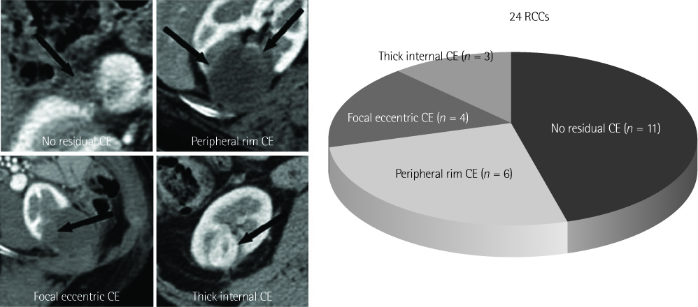
Fig. 1 The chart shows 24 renal cell carcinomas in 22 patients who underwent contrast-enhanced CT and MRI after laparoscopic renal cryoablation. At 3 month follow-up, 13 cryolesions showed residual contrast enhancement. CT images show characteristic examples of residual contrast enhancement (arrows). Note.-CE = contrast enhancement, RCC = renal cell carcinoma
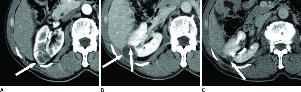
Fig. 2 Peripheral rim enhancement after cryoablation in a 73-year-old man with renal cell carcinoma. A. Contrast-enhanced CT before cryoablation shows 3.2 cm enhancing renal cell carcinoma (arrow) in the right kidney. B. Contrast-enhanced CT obtained 3 months after cryoablation shows peripheral rim enhancement (arrows) in the cryolesion. C. Contrast-enhanced CT obtained 12 months after cryoablation shows complete resolution of the peripheral rim enhancement and a reduction in the size of the cryolesion (arrow).
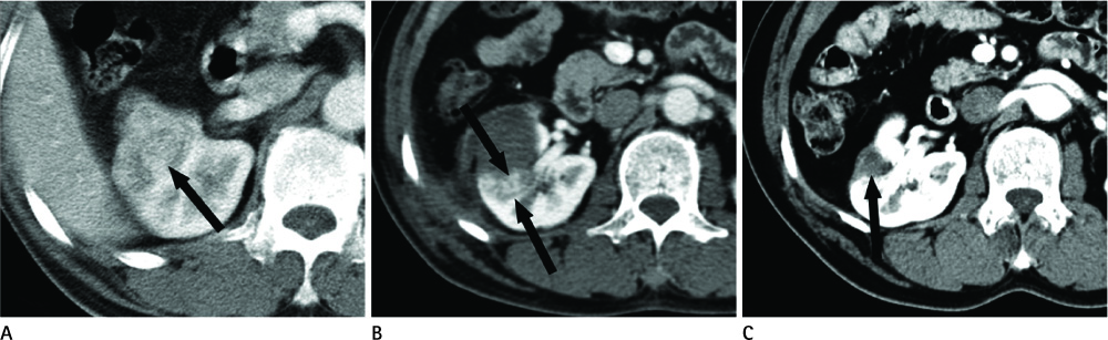
Fig. 3 Focal eccentric enhancement after cryoablation in a 52-year-old man with renal cell carcinoma. A. Contrast-enhanced CT before cryoablation shows 4.5 cm enhancing renal cell carcinoma (arrow) in the right kidney. B. Contrast-enhanced CT obtained 3 months after cryoablation shows residual focal eccentric enhancement (arrows) in the cryolesion. C. Contrast-enhanced CT obtained 18 months after cryoablation shows complete resolution of focus enhancement and a size reduction of the cryolesion (arrow).
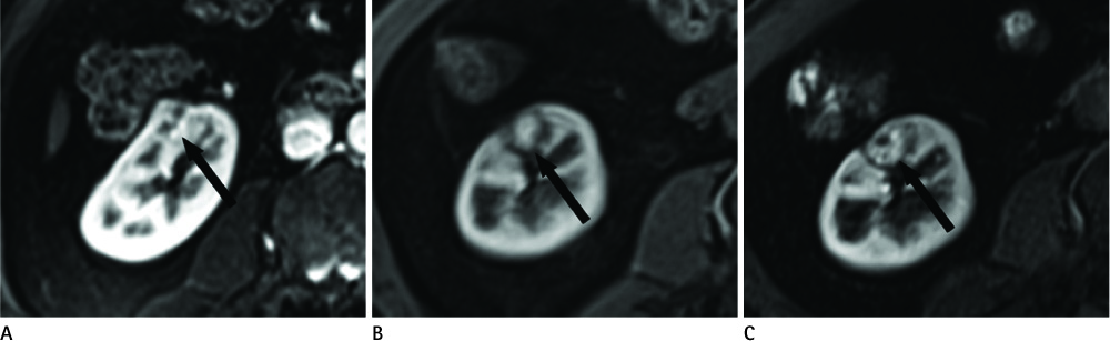
Fig. 4 Persistent thick internal enhancement after cryoablation in a 59-year-old man with renal cell carcinoma. A. Contrast-enhanced MRI before cryoablation shows 2 cm enhancing renal cell carcinoma (arrow) in the right kidney. B. Contrast-enhanced MRI obtained 3 months after cryoablation shows thick internal enhancement (arrow) in the cryolesion. C. Contrast-enhanced MRI obtained 12 months after cryoablation shows interval enlargement of the enhancing focus (arrow), representing residual viable tumor in the cryolesion.
Reference
-
1. Kaouk JH, Aron M, Rewcastle JC, Gill IS. Cryotherapy: clinical end points and their experimental foundations. Urology. 2006. 68:1 Suppl. 38–44.2. Wile GE, Leyendecker JR, Krehbiel KA, Dyer RB, Zagoria RJ. CT and MR imaging after imaging-guided thermal ablation of renal neoplasms. Radiographics. 2007. 27:325–339. discussion 339-340.3. Stein AJ, Mayes JM, Mouraviev V, Chen VH, Nelson RC, Polascik TJ. Persistent contrast enhancement several months after laparoscopic cryoablation of the small renal mass may not indicate recurrent tumor. J Endourol. 2008. 22:2433–2439.4. Porter CA 4th, Woodrum DA, Callstrom MR, Schmit GD, Misra S, Charboneau JW, et al. MRI after technically successful renal cryoablation: early contrast enhancement as a common finding. AJR Am J Roentgenol. 2010. 194:790–793.5. Patel U, Sokhi H. Imaging in the follow-up of renal cell carcinoma. AJR Am J Roentgenol. 2012. 198:1266–1276.6. Uppot RN, Silverman SG, Zagoria RJ, Tuncali K, Childs DD, Gervais DA. Imaging-guided percutaneous ablation of renal cell carcinoma: a primer of how we do it. AJR Am J Roentgenol. 2009. 192:1558–1570.7. Gervais DA, McGovern FJ, Arellano RS, McDougal WS, Mueller PR. Radiofrequency ablation of renal cell carcinoma: part 1, Indications, results, and role in patient management over a 6-year period and ablation of 100 tumors. AJR Am J Roentgenol. 2005. 185:64–71.8. Dechet CB, Zincke H, Sebo TJ, King BF, LeRoy AJ, Farrow GM, et al. Prospective analysis of computerized tomography and needle biopsy with permanent sectioning to determine the nature of solid renal masses in adults. J Urol. 2003. 169:71–74.9. Venkatesan AM, Wood BJ, Gervais DA. Percutaneous ablation in the kidney. Radiology. 2011. 261:375–391.10. Zagoria RJ. Imaging-guided radiofrequency ablation of renal masses. Radiographics. 2004. 24:Suppl 1. S59–S71.11. American College of Radiology. ACR appropriateness criteria: follow-up of renal cell carcinoma. American College of Radiology Website. 1996. Updated 2009. Accessed November 9, 2011. www.acr.org/secondarymainmenucategories/quality_safety/app_criteria/pdf/expertpanelonurologicimaging/followupofrenalcellcarcinomadoc5.aspx.12. Cestari A, Guazzoni G, dell'Acqua V, Nava L, Cardone G, Balconi G, et al. Laparoscopic cryoablation of solid renal masses: intermediate term followup. J Urol. 2004. 172(4 Pt 1):1267–1270.13. Hegarty NJ, Gill IS, Desai MM, Remer EM, O'Malley CM, Kaouk JH. Probe-ablative nephron-sparing surgery: cryoablation versus radiofrequency ablation. Urology. 2006. 68:1 Suppl. 7–13.14. Kawamoto S, Permpongkosol S, Bluemke DA, Fishman EK, Solomon SB. Sequential changes after radiofrequency ablation and cryoablation of renal neoplasms: role of CT and MR imaging. Radiographics. 2007. 27:343–355.15. Beemster P, Phoa S, Wijkstra H, de la Rosette J, Laguna P. Follow-up of renal masses after cryosurgery using computed tomography; enhancement patterns and cryolesion size. BJU Int. 2008. 101:1237–1242.16. Zhu Q, Shimizu T, Abo D, Jin M, Nagashima K, Miyasaka K. Magnetic resonance imaging findings and histopathological observations after percutaneous renal cryoablation in the rabbit model. J Urol. 2006. 175:318–326.17. Levinson AW, Su LM, Agarwal D, Sroka M, Jarrett TW, Kavoussi LR, et al. Long-term oncological and overall outcomes of percutaneous radio frequency ablation in high risk surgical patients with a solitary small renal mass. J Urol. 2008. 180:499–504. discussion 504.18. Schwartz BF, Rewcastle JC, Powell T, Whelan C, Manny T Jr, Vestal JC. Cryoablation of small peripheral renal masses: a retrospective analysis. Urology. 2006. 68:1 Suppl. 14–18.19. Zagoria RJ, Traver MA, Werle DM, Perini M, Hayasaka S, Clark PE. Oncologic efficacy of CT-guided percutaneous radiofrequency ablation of renal cell carcinomas. AJR Am J Roentgenol. 2007. 189:429–436.20. Weld KJ, Figenshau RS, Venkatesh R, Bhayani SB, Ames CD, Clayman RV, et al. Laparoscopic cryoablation for small renal masses: three-year follow-up. Urology. 2007. 69:448–451.21. Weight CJ, Kaouk JH, Hegarty NJ, Remer EM, O'Malley CM, Lane BR, et al. Correlation of radiographic imaging and histopathology following cryoablation and radio frequency ablation for renal tumors. J Urol. 2008. 179:1277–1281. discussion 1281-1283.22. Park BK, Kim CK, Lee HM. Image-guided radiofrequency ablation of Bosniak category III or IV cystic renal tumors: initial clinical experience. Eur Radiol. 2008. 18:1519–1525.23. Bolte SL, Ankem MK, Moon TD, Hedican SP, Lee FT, Sadowski EA, et al. Magnetic resonance imaging findings after laparoscopic renal cryoablation. Urology. 2006. 67:485–489.
- Full Text Links
-
- Actions
-
Cited
- CITED
-
- Close
- Share
-
- Similar articles
-
- Differentiation of Chromophobe Renal Cell Carcinoma and Clear Cell Renal Cell Carcinoma by Using Helical CT
- Magnetic resonance imaging in the diagnosis and staging of renal tumor: a comparison with computed tomography
- CT Finding of Signet Ring Cell Carcinoma of the Stomach
- Novel Experience of Contrast-Enhanced Ultrasonography to Differentiate Between Renal Cysts and Renal Cell Carcinoma
- Primary Lung Cancer: Utility of Contrast-enhanced Dynamic CT in Diagnosis with Histopathologic Correlation
