Optical Diagnosis of Small Colorectal Polyp Histology with High-Definition Colonoscopy Using Narrow Band Imaging
- Affiliations
-
- 1Division of Gastroenterology, Department of Medicine, Kansas City VA Medical Center, University of Kansas School of Medicine, Kansas City, MO, USA. amitr68@hotmail.com
- KMID: 1805293
- DOI: http://doi.org/10.5946/ce.2013.46.2.120
Abstract
- Optical diagnosis of polyp histology can potentially result in enormous cost savings by way of the "resect and discard" strategy for diminutive polyps and the "do not resect" strategy for diminutive hyperplastic polyps in the distal colon. Narrow Band Imaging (NBI) highlights the surface mucosal and vascular pattern on polyps and has been shown to accurately characterize adenomatous and hyperplastic polyps by experts. However, the results have been a little discouraging amongst lesser experienced endoscopists. Studies have also shown that using the NBI diagnosis of diminutive polyp histology, experts can accurately define the future surveillance colonoscopy intervals. However nonexperts in academic or community setting have as yet failed to achieve the recommended thresholds. The subjectivity in assessment by endoscopists leads to the variable accuracy rates and can be circumvented by computer based automated tools. Although initial experience with a few computer based algorithms have shown accuracies comparable to experts, further refinement and validation will be required before these can be implemented in clinical practice. Incorporation of optical diagnosis of diminutive polyps into clinical practice is bound to face several hurdles. But the potential for enormous cost saving makes it an attractive strategy that can make colonoscopy more cost effective.
Figure
-

Fig. 1 A) A 3-mm polyp with strong vascular pattern intensity. Histopathology showed it was an adenoma (courtesy Dr. James East, MD). (B) A 3-mm hyperplastic polyp showing weak pattern intensity (courtesy Dr. James East, MD).

Fig. 2 (A) A 2-mm hyperplastic polyp showing fine capillary network but absent mucosal pattern (bland pattern). (B) A 4-mm hyperplastic polyp showing the circular pattern with dots. (C) A 4-mm adenoma showing the round or oval pattern. (D) A 6-mm adenoma showing the tubulogyrus pattern.

Fig. 3 Lesion with clearly visible meshed capillary vessels, histologically diagnosed as an adenoma (courtesy Dr. Yasushi Sano, MD).
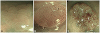
Fig. 4 (A) Capillary pattern I; hyperplastic polyp (courtesy Dr. KI Fu, MD). (B) Capillary pattern II, adenoma with low-grade dysplasia (courtesy Dr. KI Fu, MD). (C) Capillary pattern III, adenoma with high-grade dysplasia (courtesy Dr. KI Fu, MD).
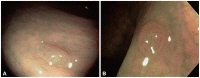
Fig. 5 (A) Hyperplastic polyp. Only very few vessels are visualized on the surface and do not show increased branching (courtesy Dr. J Tischendorf, MD). (B) Polyp showing increased density of irregular, curved and dilated blood vessels. Histologic examination showed adenoma (courtesy Dr. J Tischendorf, MD).
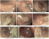
Fig. 6 Features of the narrow band imaging International Colorectal Endoscopic (NICE) criteria: (A) color, (B) vessels, and (C) surface pattern (courtesy Dr. D.K. Rex, MD).
Cited by 2 articles
-
Highlights of the 48th Seminar of Korean Society of Gastrointestinal Endoscopy
Kwang An Kwon, Il Ju Choi, Eun Young Kim, Seok Ho Dong, Ki Baik Hahm
Clin Endosc. 2013;46(3):203-211. doi: 10.5946/ce.2013.46.3.203.Diminutive and Small Colorectal Polyps: The Pathologist's Perspective
Yun Kyung Kang
Clin Endosc. 2014;47(5):404-408. doi: 10.5946/ce.2014.47.5.404.
Reference
-
1. Rex DK, Petrini JL, Baron TH, et al. Quality indicators for colonoscopy. Am J Gastroenterol. 2006; 101:873–885. PMID: 16635231.
Article2. Rastogi A, Keighley J, Singh V, et al. High accuracy of narrow band imaging without magnification for the real-time characterization of polyp histology and its comparison with high-definition white light colonoscopy: a prospective study. Am J Gastroenterol. 2009; 104:2422–2430. PMID: 19584829.
Article3. Rabeneck L, Paszat LF, Hilsden RJ, et al. Bleeding and perforation after outpatient colonoscopy and their risk factors in usual clinical practice. Gastroenterology. 2008; 135:1899–1906. PMID: 18938166.
Article4. Rex DK, Overhiser AJ, Chen SC, Cummings OW, Ulbright TM. Estimation of impact of American College of Radiology recommendations on CT colonography reporting for resection of high-risk adenoma findings. Am J Gastroenterol. 2009; 104:149–153. PMID: 19098863.
Article5. Gupta N, Bansal A, Rao D, et al. Prevalence of advanced histological features in diminutive and small colon polyps. Gastrointest Endosc. 2012; 75:1022–1030. PMID: 22405698.
Article6. Kudo S, Tamura S, Nakajima T, Yamano H, Kusaka H, Watanabe H. Diagnosis of colorectal tumorous lesions by magnifying endoscopy. Gastrointest Endosc. 1996; 44:8–14. PMID: 8836710.
Article7. Gono K, Obi T, Yamaguchi M, et al. Appearance of enhanced tissue features in narrow-band endoscopic imaging. J Biomed Opt. 2004; 9:568–577. PMID: 15189095.
Article8. Machida H, Sano Y, Hamamoto Y, et al. Narrow-band imaging in the diagnosis of colorectal mucosal lesions: a pilot study. Endoscopy. 2004; 36:1094–1098. PMID: 15578301.
Article9. Hirata M, Tanaka S, Oka S, et al. Magnifying endoscopy with narrow band imaging for diagnosis of colorectal tumors. Gastrointest Endosc. 2007; 65:988–995. PMID: 17324407.
Article10. East JE, Suzuki N, Saunders BP. Comparison of magnified pit pattern interpretation with narrow band imaging versus chromoendoscopy for diminutive colonic polyps: a pilot study. Gastrointest Endosc. 2007; 66:310–316. PMID: 17643705.
Article11. East JE, Suzuki N, Bassett P, et al. Narrow band imaging with magnification for the characterization of small and diminutive colonic polyps: pit pattern and vascular pattern intensity. Endoscopy. 2008; 40:811–817. PMID: 18828077.
Article12. Su MY, Hsu CM, Ho YP, Chen PC, Lin CJ, Chiu CT. Comparative study of conventional colonoscopy, chromoendoscopy, and narrow-band imaging systems in differential diagnosis of neoplastic and nonneoplastic colonic polyps. Am J Gastroenterol. 2006; 101:2711–2716. PMID: 17227517.
Article13. Chiu HM, Chang CY, Chen CC, et al. A prospective comparative study of narrow-band imaging, chromoendoscopy, and conventional colonoscopy in the diagnosis of colorectal neoplasia. Gut. 2007; 56:373–379. PMID: 17005766.
Article14. Rastogi A, Bansal A, Wani S, et al. Narrow-band imaging colonoscopy: a pilot feasibility study for the detection of polyps and correlation of surface patterns with polyp histologic diagnosis. Gastrointest Endosc. 2008; 67:280–286. PMID: 18155210.15. Rastogi A, Pondugula K, Bansal A, et al. Recognition of surface mucosal and vascular patterns of colon polyps by using narrow-band imaging: interobserver and intraobserver agreement and prediction of polyp histology. Gastrointest Endosc. 2009; 69(3 Pt 2):716–722. PMID: 19251016.
Article16. Rex DK. Narrow-band imaging without optical magnification for histologic analysis of colorectal polyps. Gastroenterology. 2009; 136:1174–1181. PMID: 19187781.
Article17. Sano Y, Ikematsu H, Fu KI, et al. Meshed capillary vessels by use of narrow-band imaging for differential diagnosis of small colorectal polyps. Gastrointest Endosc. 2009; 69:278–283. PMID: 18951131.
Article18. Katagiri A, Fu KI, Sano Y, et al. Narrow band imaging with magnifying colonoscopy as diagnostic tool for predicting histology of early colorectal neoplasia. Aliment Pharmacol Ther. 2008; 27:1269–1274. PMID: 18284647.
Article19. Henry ZH, Yeaton P, Shami VM, et al. Meshed capillary vessels found on narrow-band imaging without optical magnification effectively identifies colorectal neoplasia: a North American validation of the Japanese experience. Gastrointest Endosc. 2010; 72:118–126. PMID: 20381799.
Article20. Tischendorf JJ, Schirin-Sokhan R, Streetz K, et al. Value of magnifying endoscopy in classifying colorectal polyps based on vascular pattern. Endoscopy. 2010; 42:22–27. PMID: 19899031.
Article21. Hewett DG, Kaltenbach T, Sano Y, et al. Validation of a simple classification system for endoscopic diagnosis of small colorectal polyps using narrow-band imaging. Gastroenterology. 2012; 143:599–607. PMID: 22609383.
Article22. van den Broek FJ, Reitsma JB, Curvers WL, Fockens P, Dekker E. Systematic review of narrow-band imaging for the detection and differentiation of neoplastic and nonneoplastic lesions in the colon (with videos). Gastrointest Endosc. 2009; 69:124–135. PMID: 19111693.
Article23. Kobayashi Y, Hayashino Y, Jackson JL, Takagaki N, Hinotsu S, Kawakami K. Diagnostic performance of chromoendoscopy and narrow band imaging for colonic neoplasms: a meta-analysis. Colorectal Dis. 2012; 14:18–28. PMID: 20955514.
Article24. Wu L, Li Y, Li Z, Cao Y, Gao F. Diagnostic accuracy of narrow-band imaging for the differentiation of neoplastic from non-neoplastic colorectal polyps: a meta-analysis. Colorectal Dis. 2013; 15:3–11. PMID: 22251861.
Article25. Raghavendra M, Hewett DG, Rex DK. Differentiating adenomas from hyperplastic colorectal polyps: narrow-band imaging can be learned in 20 minutes. Gastrointest Endosc. 2010; 72:572–576. PMID: 20561618.
Article26. Ignjatovic A, Thomas-Gibson S, East JE, et al. Development and validation of a training module on the use of narrow-band imaging in differentiation of small adenomas from hyperplastic colorectal polyps. Gastrointest Endosc. 2011; 73:128–133. PMID: 21184878.
Article27. Kuiper T, Marsman WA, Jansen JM, et al. Accuracy for optical diagnosis of small colorectal polyps in nonacademic settings. Clin Gastroenterol Hepatol. 2012; 10:1016–1020. PMID: 22609999.
Article28. Ladabaum U, Fioritto A, Mitani A, et al. Real-time optical biopsy of colon polyps with narrow band imaging in community practice does not yet meet key thresholds for clinical decisions. Gastroenterology. 2013; 144:81–91. PMID: 23041328.
Article29. Rex DK, Kahi C, O'Brien M, et al. The American Society for Gastrointestinal Endoscopy PIVI (Preservation and Incorporation of Valuable Endoscopic Innovations) on real-time endoscopic assessment of the histology of diminutive colorectal polyps. Gastrointest Endosc. 2011; 73:419–422. PMID: 21353837.
Article30. Kessler WR, Imperiale TF, Klein RW, Wielage RC, Rex DK. A quantitative assessment of the risks and cost savings of forgoing histologic examination of diminutive polyps. Endoscopy. 2011; 43:683–691. PMID: 21623556.
Article31. Gupta N, Bansal A, Rao D, et al. Accuracy of in vivo optical diagnosis of colon polyp histology by narrow-band imaging in predicting colonoscopy surveillance intervals. Gastrointest Endosc. 2012; 75:494–502. PMID: 22032847.
Article32. Hewett DG, Huffman ME, Rex DK. Leaving distal colorectal hyperplastic polyps in place can be achieved with high accuracy by using narrow-band imaging: an observational study. Gastrointest Endosc. 2012; 76:374–380. PMID: 22695207.
Article33. Paggi S, Rondonotti E, Amato A, et al. Resect and discard strategy in clinical practice: a prospective cohort study. Endoscopy. 2012; 44:899–904. PMID: 22859259.
Article34. Gross S, Trautwein C, Behrens A, et al. Computer-based classification of small colorectal polyps by using narrow-band imaging with optical magnification. Gastrointest Endosc. 2011; 74:1354–1359. PMID: 22000791.
Article35. Takemura Y, Yoshida S, Tanaka S, et al. Computer-aided system for predicting the histology of colorectal tumors by using narrow-band imaging magnifying colonoscopy (with video). Gastrointest Endosc. 2012; 75:179–185. PMID: 22196816.
Article36. Rex DK, Fennerty MB, Sharma P, Kaltenbach T, Soetikno R. Bringing new endoscopic imaging technology into everyday practice: what is the role of professional GI societies? Polyp imaging as a template for moving endoscopic innovation forward to answer key clinical questions. Gastrointest Endosc. 2010; 71:142–146. PMID: 19922926.
Article
- Full Text Links
-
- Actions
-
Cited
- CITED
-
- Close
- Share
-
- Similar articles
-
- Introduction: What Are New Roles of Current Colonoscopy?
- Optical Diagnosis for Colorectal Polyps: A Useful Technique Now or in the Future?
- Artificial Intelligence-Based Colorectal Polyp Histology Prediction by Using Narrow-Band Image-Magnifying Colonoscopy
- Polyp Detection, Characterization, and Management Using Narrow-Band Imaging with/without Magnification
- Optical diagnosis by near-focus versus normal-focus narrow band imaging colonoscopy in colorectal polyps based on combined NICE and WASP classification: a randomized controlled trial
