Consolidations in Nodular Bronchiectatic Mycobacterium Avium Complex Lung Disease: Mycobacterium Avium Complex or Other Infection?
- Affiliations
-
- 1Department of Radiology, Seoul St. Mary's Hospital, The Catholic University of Korea, Seoul, Korea. sghnk@catholic.ac.kr
- KMID: 1805192
- DOI: http://doi.org/10.3349/ymj.2010.51.4.546
Abstract
- PURPOSE
The objective of this study is to define the clinical implications of consolidations in nodular bronchiectatic type Mycobacterium avium complex (NB-MAC) infection.
MATERIALS AND METHODS
A total of 69 patients (M : F = 17 : 52; mean age, 64 years; age range, 41-85 years) with MAC isolated in the sputum culture and nodular bronchiectasis on the initial and follow-up CT scans were included. We retrospectively reviewed the incidence of consolidation and analyzed its clinical course by using radiographic changes with or without anti-MAC drug therapy.
RESULTS
In 44 of the 69 cases (64%), focal consolidations were seen on the initial and follow-up CT images. In 35 of the 44 (80%) cases, consolidations completely regressed, and in 3 cases (7%), consolidations partially regressed within 2 months with only antibiotics. In 2 cases (5%), the consolidations remained stable for over 2 months without anti-MAC drug therapy. Only in 4 cases (9%) did the consolidations improve after anti-MAC drug therapy. In 11 of the 38 cases (29%) with responsiveness to antibiotics, non-mycobacterial micro-organisms were identified in sputum, including pseudomonas, hemophilus, staphylococcus, and others.
CONCLUSION
In NB-MAC, consolidations are commonly present on CT. In these conditions, most of consolidations result from pneumonia other than MAC.
Figure
-
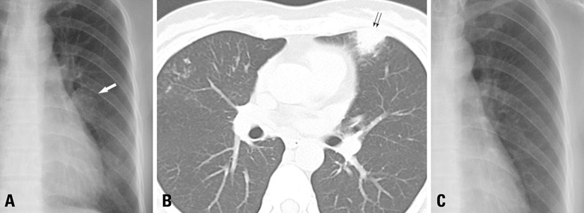
Fig. 1 A 45-year-old woman with NB-MAC, initially presenting with consolidation. (A) Plain chest radiography shows an ill-defined ovoid consolidation in left middle lung zone (arrow). (B) Initial CT scans shows mild cylindrical bronchiectasis and adjacent centrilobular nodules in right middle lobe and lingula, suggestive of NB-MAC. A focal consolidation is seen in subpleural region of lingula (double arrows). (C) FU plain radiography after 2 weeks shows disappearance of the prior focal consolidation after antibiotic therapy. NB-MAC, nodular bronchiectatic type Mycobacterium avium complex; FU, follow-up.

Fig. 2 A 48-year-old man with NB-MAC, who had a consolidation without response to antibiotics. (A and B) Initial CT shows multifocal clusters of centrilobular nodules with tree in bud appearance in right upper and middle lobes and lingula (arrowheads). Peripheral airways of right upper lobe are minimally dilated (thin arrow). A focal consolidation is seen at anterior subpleural region of lingula (arrow). (C) Two-month FU CT shows little change in size of the prior consolidation in lingula (double arrows) after empirical antibiotics. NB-MAC, nodular bronchiectatic type Mycobacterium avium complex; FU, follow-up.

Fig. 3 A 53-year-old women with NB-MAC, who had newly detected consolidations during FU period managed with empirical antibiotic therapy. (A) Initial CT shows bronchiectasis with atelectasis and nearby clustered micronodules in right middle lobe. Faint nodular opacities are also visible at posterior aspect of right lower lobe (arrowheads). (B) Six-month FU HRCT at the same level to A shows a new irregular consolidation at posterior subpleural region of right lower lobe (arrow). Another smaller consolidation is seen in right middle lobe (double arrows). Bronchiectasis and clustered centrilobular nodules of right middle lobe, indicative of NB-MAC, are more clearly demarcated than before. The pre-existing faint nodules of right lower lobe are invisible, suggestive of improvement of focal bronchiolitis. (C) One-month FU HRCT after antibiotic therapy shows complete regression of the prior consolidations in right middle and lower lobes. NB-MAC, nodular bronchiectatic type Mycobacterium avium complex; FU, follow-up; HRCT, high-resolution CT.

Fig. 4 A 72-year-old man with NB-MAC, who had newly detected consolidations during FU with response to anti-MAC therapy. (A) Axial CT shows multifocal cylindrical bronchiectasis and centrilobular nodules with volume loss in both lungs. Irregular consolidations are seen at posterior aspect of right lower lobe (arrow). Anti-MAC therapy was initiated. (B) Plain radiograph obtained at the same day to A shows multifocal consolidations and small nodular opacities in right lung and left lower lung zone. (C) FU plain radiography after 2 months shows decreased extent of the consolidations and nodules of both lungs. NB-MAC, nodular bronchiectatic type Mycobacterium avium complex; FU, follow-up.
Reference
-
1. Diagnosis and treatment of disease caused by nontuberculous mycobacteria. This official statement of the American Thoracic Society was approved by the Board of Directors, March 1997. Medical Section of the American Lung Association. Am J Respir Crit Care Med. 1997. 156:S1–S25.2. Koh WJ, Kwon OJ, Lee KS. Nontuberculous mycobacterial pulmonary diseases in immunocompetent patients. Korean J Radiol. 2002. 3:145–157.3. O'Brien RJ, Geiter LJ, Snider DE Jr. The epidemiology of nontuberculous mycobacterial diseases in the United States. Results from a national survey. Am Rev Respir Dis. 1987. 135:1007–1014.4. Tsukamura M, Kita N, Shimoide H, Arakawa H, Kuze A. Studies on the epidemiology of nontuberculous mycobacteriosis in Japan. Am Rev Respir Dis. 1988. 137:1280–1284.5. Prince DS, Peterson DD, Steiner RM, Gottlieb JE, Scott R, Israel HL, et al. Infection with Mycobacterium avium complex in patients without predisposing conditions. N Engl J Med. 1989. 321:863–868.6. Reich JM, Johnson RE. Mycobacterium avium complex pulmonary disease presenting as an isolated lingular or middle lobe pattern. The Lady Windermere syndrome. Chest. 1992. 101:1605–1609.7. Koh WJ, Lee KS, Kwon OJ, Jeong YJ, Kwak SH, Kim TS. Bilateral bronchiectasis and bronchiolitis at thin-section CT: diagnostic implications in nontuberculous mycobacterial pulmonary infection. Radiology. 2005. 235:282–288.
Article8. Jeong YJ, Lee KS, Koh WJ, Han J, Kim TS, Kwon OJ. Nontuberculous mycobacterial pulmonary infection in immunocompetent patients: comparison of thin-section CT and histopathologic findings. Radiology. 2004. 231:880–886.
Article9. Kim TS, Koh WJ, Han J, Chung MJ, Lee JH, Lee KS, et al. Hypothesis on the evolution of cavitary lesions in nontuberculous mycobacterial pulmonary infection: thin-section CT and histopathologic correlation. AJR Am J Roentgenol. 2005. 184:1247–1252.10. Fujita J, Ohtsuki Y, Shigeto E, Suemitsu I, Yamadori I, Bandoh S, et al. Pathological findings of bronchiectases caused by Mycobacterium avium intracellulare complex. Respir Med. 2003. 97:933–938.11. Fujita J, Ohtsuki Y, Suemitsu I, Shigeto E, Yamadori I, Obayashi Y, et al. Pathological and radiological changes in resected lung specimens in Mycobacterium avium intracellulare complex disease. Eur Respir J. 1999. 13:535–540.12. Austin JH, Müller NL, Friedman PJ, Hansell DM, Naidich DP, Remy-Jardin M, et al. Glossary of terms for CT of the lungs: recommendations of the Nomenclature Committee of the Fleischner Society. Radiology. 1996. 200:327–331.
Article13. Wickremasinghe M, Ozerovitch LJ, Davies G, Wodehouse T, Chadwick MV, Abdallah S, et al. Non-tuberculous mycobacteria in patients with bronchiectasis. Thorax. 2005. 60:1045–1051.14. Okumura M, Iwai K, Ogata H, Ueyama M, Kubota M, Aoki M, et al. Clinical factors on cavitary and nodular bronchiectatic types in pulmonary Mycobacterium avium complex disease. Intern Med. 2008. 47:1465–1472.15. Fujiuchi S, Matsumoto H, Yamazaki Y, Nakao S, Takahashi M, Satoh K, et al. Analysis of chest CT in patients with Mycobacterium avium complex pulmonary disease. Respiration. 2003. 70:76–81.
Article
- Full Text Links
-
- Actions
-
Cited
- CITED
-
- Close
- Share
-
- Similar articles
-
- Rapid identification of mycobacterium avium and mycobacterium intracellulare by the amplification of rRNA sequences
- Respiratory Review of 2009: Nontuberculous Mycobacterium
- A Case of Pulmonary and Endobronchial Mycobacterium avium Infection Presenting as an Acute Pneumonia in an Immunocompetent Patient
- Vertebral Osteomyelitis due to Mycobacterium intracellulare in an Immunocompetent Elderly Patient After Vertebroplasty
- Disseminated Mycobacterium avium Complex Infection in a Patient with Acquired Immunodeficiency Syndrome
