Primary Leiomyoma of Ureter Coexisting with Renal Cell Carcinoma: A Case Report
- Affiliations
-
- 1Department of Radiology, Eulji University Hospital, Daejeon, Korea. orionphil@hotmail.com
- KMID: 1801556
- DOI: http://doi.org/10.3348/jksr.2014.71.6.320
Abstract
- Mesenchymal origin of ureter tumors account for less than 3 percent of all primary ureteral tumors. Among mesenchymal tumors, primary leiomyoma of ureter is extremely rare. Here, we present a case of primary leiomyoma of ureter coexisting with renal cell carcinoma. When encountering well-defined homogeneously enhanced mass of ureter on computed tomography, radiologist should keep in mind that ureteral leiomyoma should be considered as differential diagnosis.
Figure
-
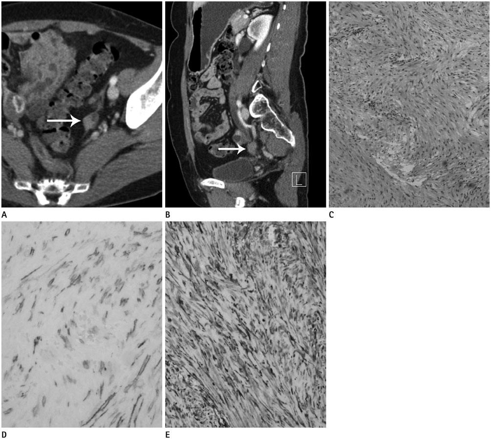
Fig. 1 50-year-old woman with primary leiomyoma of the ureter. A. Contrast-enhanced abdomen CT shows well-defined small homogeneously enhancing mass (about 1.4 × 1.1 cm) at the medial aspect of the left distal ureter. The periureteral mass (arrow) was compressing the left distal ureter. B. Contrast-enhanced abdomen CT shows obstructive hydronephrosis and hydroureter of the left kidney (arrow) and slightly decreased enhancement of renal parenchyma suggesting obstructive uropathy. C. Microscopic image of the mass shows well-differentiated multiple layers of hypercellular and hyperchromatic interwoven bundles of smooth muscle cells (H&E, × 40). D, E. On the immunohistochemical staining, the spindle cells were positive for the smooth muscle actin (SMA) (D) and vimentin (E) (SMA, × 100, vimentin, × 100).

Fig. 2 Coexistent renal cell carcinoma in the mid-portion of the left kidney. A, B. Contrast-enhanced abdomen CT with axial scan shows 1.4 × 1.3 cm sized hypervascular mass (arrow) in the mid-portion of left renal parenchyma. This mass showed hyperenhancement on corticomedullary phase (A) and washout of enhancement on excretory phase (B). It was considered as a renal cell carcinoma. C. Micrograph shows epithelial cells with clear cytoplasm and a distinct cell membrane, separated by a delicate branching network of vascular tissue (H&E, × 100). It is typical histologic findings of renal clear-cell carcinoma.
Reference
-
1. Alvarado-Cabrero I, Candanedo-González F, Sosa-Romero A. Leiomyoma of the urethra in a Mexican woman: a rare neoplasm associated with the expression of estrogen receptors by immunohistochemistry. Arch Med Res. 2001; 32:88–90.2. Kurokawa S, Kojima Y, Tozawa K, Hayashi Y, Sasaki S, Kohri K. Female paraurethral leiomyoma: immunohistochemical approach to the relationship between leiomyoma and ovarian hormones. J Urol. 2002; 167:1403–1404.3. Mondschein LJ, Sutton AP, Rothfeld SH. Leiomyoma of the ureter in a child: the first reported case. J Urol. 1976; 116:516–518.4. Werth DD, Weigel JW, Mebust WK. Primary neoplasms of the ureter. J Urol. 1981; 125:628–631.5. Zehri AA, Ali A, Iqbal F, Jessca M. Primary leiomyoma--a rare tumour of ureter. J Pak Med Assoc. 2013; 63:268–270.6. Alam NA, Olpin S, Rowan A, Kelsell D, Leigh IM, Tomlinson IP, et al. Missense mutations in fumarate hydratase in multiple cutaneous and uterine leiomyomatosis and renal cell cancer. J Mol Diagn. 2005; 7:437–443.7. Nouralizadeh A, Tabibi A, Mahmoudnejad N, Taheri M, Torbati PM. Partial ureterectomy for a huge primary leiomyoma of the ureter. J Pak Med Assoc. 2010; 60:62–64.8. Casillas J, Joseph RC, Guerra JJ Jr. CT appearance of uterine leiomyomas. Radiographics. 1990; 10:999–1007.
- Full Text Links
-
- Actions
-
Cited
- CITED
-
- Close
- Share
-
- Similar articles
-
- Primary Squamous Cell Carcinoma of the Endometrium Covering Submucosal Leiomyoma
- Primary Squamous Cell Carcinoma of the Ureter after Reterocutaneoustomy
- Bilateral primary carcinoma of the ureter associated with acute renal failure
- Primary Squamous Cell Carcinoma of the Ureter
- A Case or Squamous Cell Carcinoma of the Renal Pelvis Associated with Squamous Cell Carcinoma in Situ of the Ureter
