Ann Surg Treat Res.
2015 Jun;88(6):306-310. 10.4174/astr.2015.88.6.306.
In vivo porcine training model for laparoscopic Roux-en-Y choledochojejunostomy
- Affiliations
-
- 1Department of Surgery, Seoul St. Mary's Hospital, The Catholic University of Korea College of Medicine, Seoul, Korea. gshth@catholic.ac.kr
- KMID: 1800643
- DOI: http://doi.org/10.4174/astr.2015.88.6.306
Abstract
- PURPOSE
The purpose of this study was to develop a porcine training model for laparoscopic choledochojejunostomy (CJ) that can act as a bridge between simulation models and actual surgery for novice surgeons. The feasibility of this model was evaluated.
METHODS
Laparoscopic CJ using intracorporeal sutures was performed on ten animals by a surgical fellow with no experience in human laparoscopic CJ. A single layer of running sutures was placed in the posterior and anterior layers. Jejunojejunostomy was performed using a linear stapler, and the jejunal opening was closed using absorbable unidirectional sutures (V-Loc 180).
RESULTS
The average operation time was 131.3 +/- 36.4 minutes, and the CJ time was 57.5 +/- 18.4 minutes. Both the operation time and CJ time showed a steady decrease with an increasing number of cases. The average diameter of the CBD was 6.4 +/- 0.8 mm. Of a total of ten animals, eight were sacrificed after the procedure. In two animals, a survival model was evaluated. Both pigs recovered completely and survived for two weeks, after which both animals were sacrificed. None of the animals exhibited any signs of bile leakage or anastomosis site stricture.
CONCLUSION
The porcine training model introduced in this paper is an adequate model for practicing laparoscopic CJ. Human tissue simulation is excellent.
Keyword
Figure
-
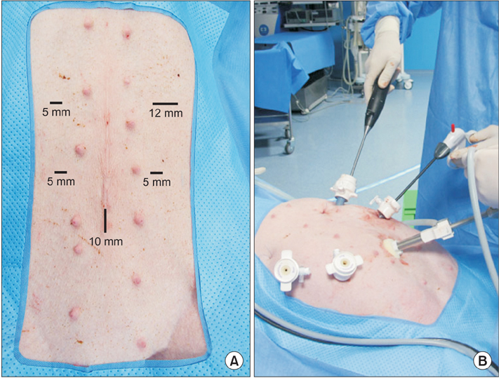
Fig. 1 Trocar placement and surgeon position during laparoscopic choledochojejunostomy (CJ). (A) Trocar placement. (B) Position of the surgeon during laparoscopic CJ.
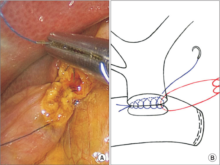
Fig. 2 (A) Posterior wall suture of choledochojejunostomy. (B) Schematic illustration.
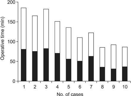
Fig. 3 Operative time by case number. Black bar: choledochojejunostomy time.
Reference
-
1. Han HS, Yi NJ. Laparoscopic Roux-en-Y choledochojejunostomy for benign biliary disease. Surg Laparosc Endosc Percutan Tech. 2004; 14:80–84.2. Gumbs AA, Hoffman JP. Laparoscopic radical cholecystectomy and Roux-en-Y choledochojejunostomy for gallbladder cancer. Surg Endosc. 2010; 24:1766–1768.3. Reznick RK, MacRae H. Teaching surgical skills: changes in the wind. N Engl J Med. 2006; 355:2664–2669.4. Hon SS, Ng SS, Lee JF, Li JC, Lo AW. In vitro porcine training model for colonic endoscopic submucosal dissection: an inexpensive and safe way to acquire a complex endoscopic technique. Surg Endosc. 2010; 24:2439–2443.5. Narumi S, Toyoki Y, Ishido K, Kudo D, Umehara M, Kimura N, et al. Introduction of a simulation model for choledochoand pancreaticojejunostomy. Hepatogastroenterology. 2012; 59:2333–2334.6. Bauer F, Rommel N, Kreutzer K, Weitz J, Wagenpfeil S, Gulati A, et al. A novel approach to teaching surgical skills to medical students using an ex vivo animal training model. J Surg Educ. 2014; 71:459–465.7. Jeong SH, Park JH, Yoo MW, Choi SK, Hong SC, Jung EJ, et al. Feasibility of the transumbilical route compared with the transoral route in gastric upper body endoscopic submucosal dissection: a porcine model. Surg Endosc. 2014; 28:515–523.8. Milsom JW, Trencheva K, Ezell P, Maggiori L, Pavoor R, Vitellaro M, et al. Feasibility and safety of laparoscopic colon surgery performed under intravenous sedation and local anesthesia using microinvasive (<3 mm) instruments: an acute and survival study on porcine model. Surg Innov. 2015; 22:131–136.9. Jayaraman S, Quan D, Al-Ghamdi I, El-Deen F, Schlachta CM. Does robotic assistance improve efficiency in performing complex minimally invasive surgical procedures? Surg Endosc. 2010; 24:584–588.10. Schob OM, Day PW, Josloff RK, Zucker KA. An experimental teaching model for laparoscopic choledochojejunostomy. Surg Laparosc Endosc. 1996; 6:341–347.11. Reed DN Jr, Cacchione RN, Allen JW, Arlauskas V, Casey J, Larson GM, et al. Laparoscopic choledochojejunostomy and gastrojejunostomy in a porcine model. Surg Endosc. 2003; 17:86–88.12. Suzuki O, Hirano S, Yano T, Okamura K, Hazama K, Shichinohe T, et al. Laparoscopic pancreaticoduodenectomy is effective in a porcine model. Surg Endosc. 2008; 22:2509–2513.13. Laursen HB, Thorsoe HJ, Funch-Jensen P, Rokkjaer M, Yasuda Y, Mortensen FV. Robotic-assisted laparoscopic Roux-en-Y choledochojejunostomy in pigs. J Hepatobiliary Pancreat Surg. 2005; 12:167–172.14. Ruurda JP, van Dongen KW, Dries J, Borel Rinkes IH, Broeders IA. Robot-assisted laparoscopic choledochojejunostomy. Surg Endosc. 2003; 17:1937–1942.
- Full Text Links
-
- Actions
-
Cited
- CITED
-
- Close
- Share
-
- Similar articles
-
- The rat choledochojejunostomy model for microsurgical training
- In vivo porcine training model of laparoscopic common bile duct repair with T-tube insertion under the situation of iatrogenic common bile duct injury
- The Impact of Using a Porcine Model in Laparoscopic Partial Nephrectomy Training
- Robotic Roux-en-Y Gastric Bypass and Robotic Sleeve Gastrectomy for Morbid Obesity: Case Reports
- Laparoscopic Hiatal Hernia Repair and Roux-en-Y Conversion for Refractory Duodenogastroesophageal Reflux after Billroth I Distal Gastrectomy
