Clear Cell Chondrosarcoma of the Tibia Diaphysis: A Case Report
- Affiliations
-
- 1Department of Orthopedic Surgery, School of Medicine, Kyung Hee University, Seoul, Korea. cshan1129@yahoo.co.kr
- KMID: 1799899
- DOI: http://doi.org/10.5292/jkbjts.2014.20.2.89
Abstract
- Clear cell chondrosarcoma is a very rare malignant bone tumor that shows a strong predilection for the epiphysis or metaphysis of long bones. Many studies have reported that the proximal end of the femur is the most commonly affected site, followed by the proximal end of the humerus. Histopathologically, tumor cells of this type have centrally located round nucleoli with clear cytoplasm and a distinct cytoplasmic membrane. Generally, clear cell chondrosarcomas is not confused with conventional chondrosarcomas. However, when it involves the diaphysis in long bones, diagnosis can be hindered, as only three reports of this exist in the literature. We report herein an unusual case of clear cell chondrosarcoma of the tibial diaphysis in a 42-year-old male.
Keyword
MeSH Terms
Figure
-
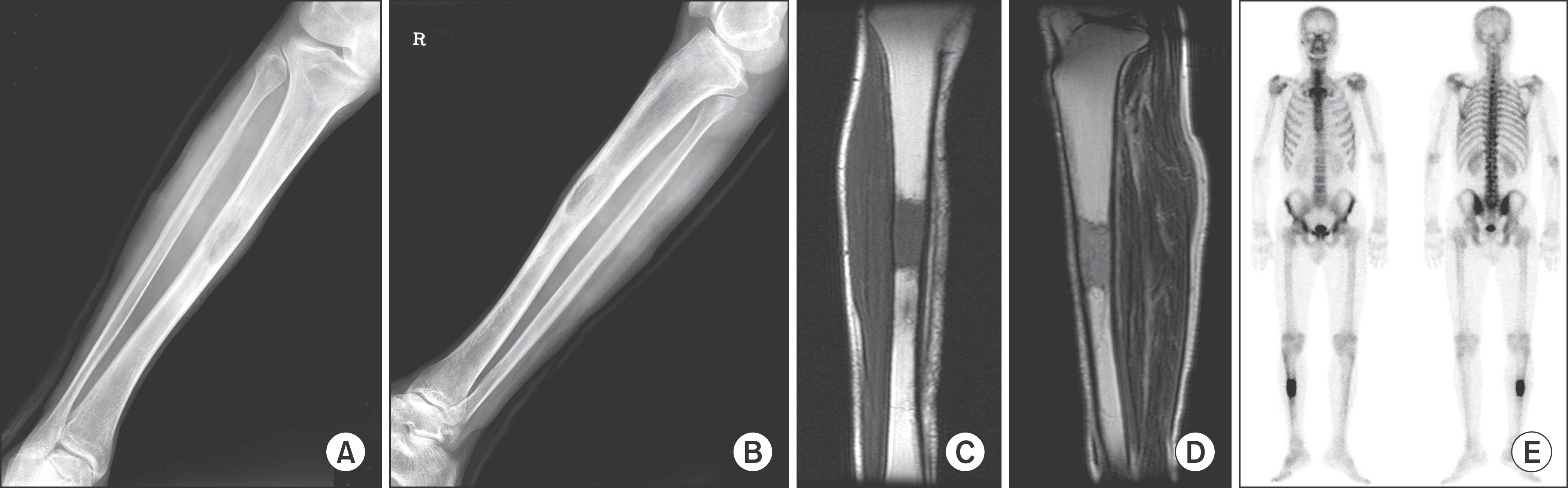
Figure 1. (A, B) The radiographs showed an intramedullary ovalshaped radiolucent lesion of the right tibia diaphysis. A distinct sclerotic border was identified, but the posterior endosteal portion of the lesion showed an ill-defined margin with a slightly infiltrative pattern. There was no periosteal reaction. (C) T1-weighted coronal images showed a well-marginated lesion with intermediate signal intensity in the medullary cavity of the tibia shaft. (D) T2-weighted sagittal images showed intermediate to high signal intensity with a relatively homogeneous appearance without soft tissue expansion. (E) A bone scan revealed highly increased uptake in the middle shaft of the tibia, and showed no other abnormalities elsewhere.
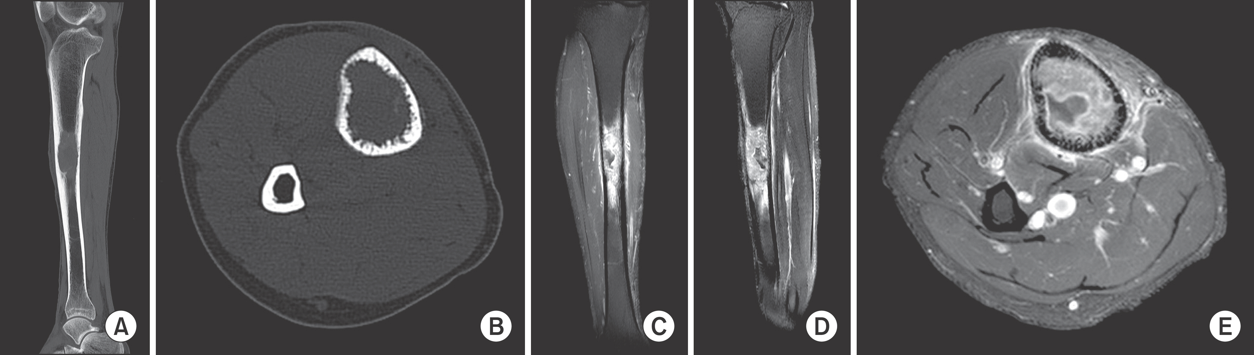
Figure 2. (A, B) CT showed an intramedullary lesion with permeative endosteal bone destruction. Complete cortical disruption was not noted. (C−E) MRI repeated after surgical biopsy showed a similar appearance with enhancement.
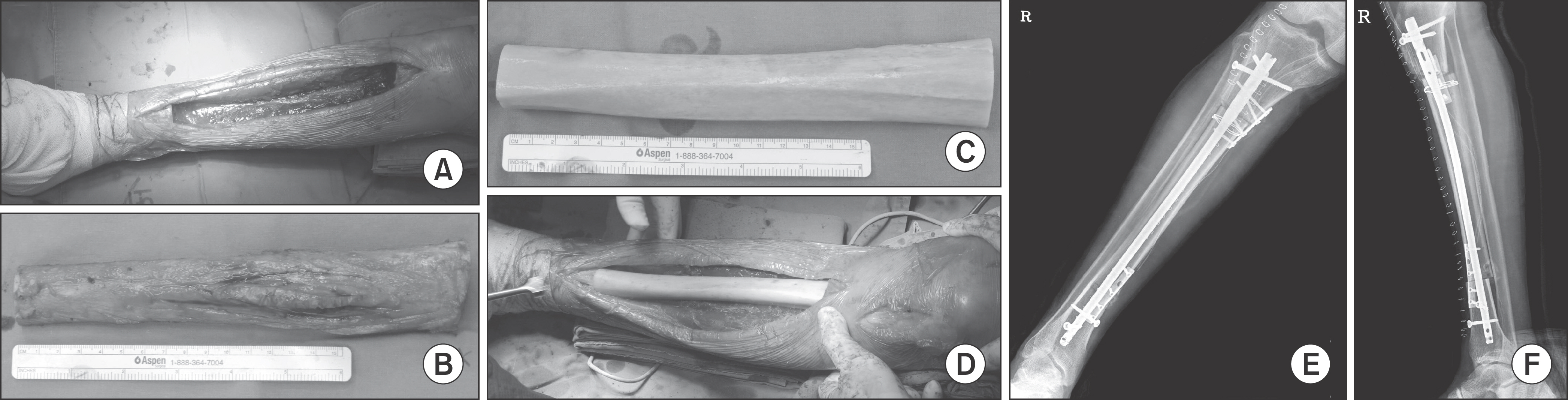
Figure 3. (A−F) Resection of the tibia shaft including the tumor was performed and structural allogenic bone was transplanted into the empty space. The allo-bone was fixed with an intramedullary nail and plate.

Figure 4. Histopathologic appearance. The cells had pale, clear cytoplasm with a distinct cytoplasmic membrane and centrally located large, round nuclei, which are typical features of CCCS (H&E, ×160, ×250).
Cited by 1 articles
-
Clear Cell Chondrosarcoma Mimicking Benign Bone Tumors
Chang-Bae Kong, Seung Yong Lee, Won-Seok Song, Wan-Hyeong Cho, Jae-Soo Koh, Dae-Geun Jeon
J Korean Orthop Assoc. 2018;53(1):51-57. doi: 10.4055/jkoa.2018.53.1.51.
Reference
-
References
1. Unni KK, Dahlin DC, Beabout JW, Sim FH. Chondrosarcoma: clear-cell variant. A report of sixteen cases. J Bone Joint Surg Am. 1976; 58:676–83.2. Bjornsson J, Unni KK, Dahlin DC, Beabout JW, Sim FH. Clear cell chondrosarcoma of bone. Observations in 47 cases. Am J Surg Pathol. 1984; 8:223–30.3. Memis A, Arkun R, Basdemir G, Sabah D, Ustün EE. Clear cell chondrosarcoma: unusual radiologic appearances with histologic correlation. Eur Radiol. 2002; 12:427–30.
Article4. Itälä A, Leerapun T, Inwards C, Collins M, Scully SP. An institutional review of clear cell chondrosarcoma. Clin Orthop Relat Res. 2005; 440:209–12.5. Giuffrida AY, Burgueno JE, Koniaris LG, Gutierrez JC, Duncan R, Scully SP. Chondrosarcoma in the United States (1973 to 2003): an analysis of 2890 cases from the SEER database. J Bone Joint Surg Am. 2009; 91:1063–72.
Article6. Collins MS, Koyama T, Swee RG, Inwards CY. Clear cell chondrosarcoma: radiographic, computed tomographic, and magnetic resonance findings in 34 patients with pathologic correlation. Skeletal Radiol. 2003; 32:687–94.
Article7. Ayoub KS, Grimer RJ, Carter SR, Mangham DC, Davies AM, Tillman RM. Clear cell chondrosarcoma of bone. Sarcoma. 1999; 3:115–9.
Article8. Present D, Bacchini P, Pignatti G, Picci P, Bertoni F, Campanacci M. Clear cell chondrosarcoma of bone. A report of 8 cases. Skeletal Radiol. 1991; 20:187–91.9. Unni KK, Dahlin DC, McLeod RA, Pritchard DJ. Intraosseous well-differentiated osteosarcoma. Cancer. 1977; 40:1337–47.
Article10. Andresen KJ, Sundaram M, Unni KK, Sim FH. Imaging features of low-grade central osteosarcoma of the long bones and pelvis. Skeletal Radiol. 2004; 33:373–9.
Article
