Imaging Sci Dent.
2014 Jun;44(2):129-135. 10.5624/isd.2014.44.2.129.
Assessment of trabecular bone changes around endosseous implants using image analysis techniques: A preliminary study
- Affiliations
-
- 1Department of Oral Medicine and Radiology, Benghazi University College of Dentistry, Benghazi, Libya.
- 2Oral Diagnosis and Polyclinics, Faculty of Dentistry, The University of Hong Kong, Hong Kong. jellodent@yahoo.com
- 3Department of Oral Radiology, University Dental Hospital of Manchester, Manchester, United Kingdom.
- KMID: 1799594
- DOI: http://doi.org/10.5624/isd.2014.44.2.129
Abstract
- PURPOSE
The objective of this study was to assess the trabecular bone changes that occurred around functional endosseous dental implants by means of radiographic image analysis techniques.
MATERIALS AND METHODS
Immediate preoperative and postoperative periapical radiographs of de-identified implant patients at the University Dental Hospital of Manchester were retrieved, screened for specific inclusion criteria, digitized, and quantified for structural elements of the trabecular bone around the endosseous implants, by using image analysis techniques. Data were analyzed using SPSS version 11.5. P values of less than 0.05 were considered statistically significant.
RESULTS
A total of 12 implants from 11 patients were selected for the study, and 26 regions of interest were obtained. There was a significant increase in the bone area in terms of the mean distance between nodes (p=0.006) and a significant decrease in the marrow area in terms of the bone area (p=0.006) and the length of marrow spaces (p=0.032).
CONCLUSION
It appeared that the bone around the implant underwent remodeling that resulted in a net increase in bone after implant placement.
MeSH Terms
Figure
-

Fig. 1 Digitization, selection, and cropping of the images. (a) A periapical radiograph is digitized by a scanner using an 8-bit gray scale and a resolution of 1200 dpi. (b) A magnified region of interest (ROI) of 150×150 pixels in the tagged image file (TIF) format below the root apex is cropped by Adobe Photoshop Elements version 3. (c) A magnified ROI of 150×75 pixels in the TIF at the mesial aspect of the root apex. (d) A magnified ROI of 150×75 pixels in the TIF format at the distal aspect of the root apex.
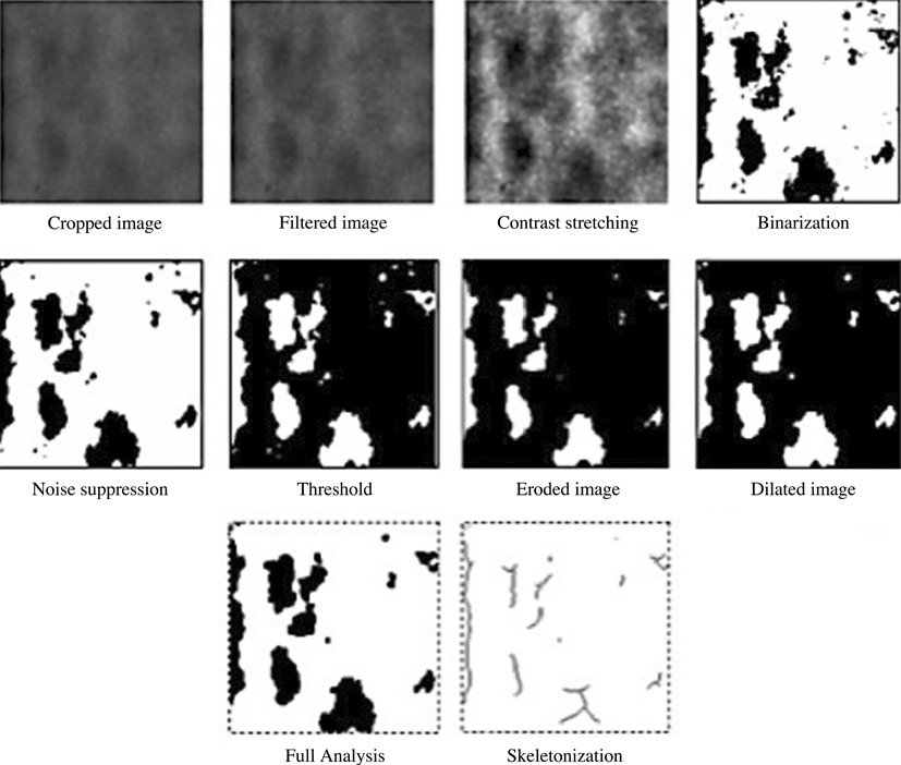
Fig. 2 The images are the procedures for image digitization, optimization, and analysis (marrow).

Fig. 3 Inverted image is analyzed (trabecular) using Win TAS for the images shown in Fig. 2.
Reference
-
1. Brägger U. Radiographic parameters for the evaluation of peri-implant tissues. Periodontol 2000. 1994; 4:87–97.
Article2. Moy PK, Medina D, Shetty V, Aghaloo TL. Dental implant failure rates and associated risk factors. Int J Oral Maxillofac Implants. 2005; 20:569–577.3. Miles DA, Razzano MR. The future of digital imaging in dentistry. Dent Clin North Am. 2000; 44:427–438.4. White SC, Rudolph DJ. Alterations of the trabecular pattern of the jaws in patients with osteoporosis. Oral Surg Oral Med Oral Pathol Oral Radiol Endod. 1999; 88:628–635.
Article5. White SC. Oral radiographic predictors of osteoporosis. Dentomaxillofac Radiol. 2002; 31:84–92.
Article6. Aaron JE, Shore PA. Histomorphometry. In : Langton CM, Njeh CF, editors. The physical measurement of bone. 2nd ed. Bristol, Philadelphia: Institute of Physics Pub;2004. p. 185–224.7. Chappard D, Guggenbuhl P, Legrand E, Baslé MF, Audran M. Texture analysis of X-ray radiographs is correlated with bone histomorphometry. J Bone Miner Metab. 2005; 23:24–29.
Article8. Ludlow JB, Gates W, Nason R Jr. Radiographic evaluation of implant-obscured bone. Comparison of digitally subtracted tomographic and periapical techniques. Oral Surg Oral Med Oral Pathol Oral Radiol Endod. 1995; 80:351–357.9. Jonasson G, Bankvall G, Kiliaridis S. Estimation of skeletal bone mineral density by means of the trabecular pattern of the alveolar bone, its interdental thickness, and the bone mass of the mandible. Oral Surg Oral Med Oral Pathol Oral Radiol Endod. 2001; 92:346–352.
Article10. Gröndahl K, Sundén S, Gröndahl HG. Inter- and intraobserver variability in radiographic bone level assessment at Brånemark fixtures. Clin Oral Implants Res. 1998; 9:243–250.
Article11. Benn DK. A review of the reliability of radiographic measurements in estimating alveolar bone changes. J Clin Periodontol. 1990; 17:14–21.
Article12. Leonhardt A, Gröndahl K, Bergström C, Lekholm U. Long-term follow-up of osseointegrated titanium implants using clinical, radiographic and microbiological parameters. Clin Oral Implants Res. 2002; 13:127–132.
Article13. Borg E, Gröndahl K, Persson LG, Gröndahl HG. Marginal bone level around implants assessed in digital and film radiographs: in vivo study in the dog. Clin Implant Dent Relat Res. 2000; 2:10–17.
Article14. Wennström JL, Ekestubbe A, Gröndahl K, Karlsson S, Lindhe J. Implant-supported single-tooth restorations: a 5-year prospective study. J Clin Periodontol. 2005; 32:567–574.
Article15. Frost HM. Wolff's Law and bone's structural adaptations to mechanical usage: an overview for clinicians. Angle Orthod. 1994; 64:175–188.
- Full Text Links
-
- Actions
-
Cited
- CITED
-
- Close
- Share
-
- Similar articles
-
- Bone graft procedure with endosseous implants : A review of the literature
- THREE-DIMENSIONAL FINI6E ELEMENT ANALYSIS OF THE ENDOSSEOUS IMPLANT DESIGNS
- Changes in the fractal dimension of peri-implant trabecular bone after loading: a retrospective study
- CLINICAL STUDY OF ENDOSSEOUS HYDROXYAPATITE COATED IMPLANTS
- Clinical evaluation of prognosis of osseointegrated dental implant in treatment of maxillary edentulous area
