Development of dental charts according to tooth development and eruption for Turkish children and young adults
- Affiliations
-
- 1University of Istanbul, Cerrahpasa Faculty of Medicine, Forensic Medicine Department, Istanbul, Turkey. bkaradayi1970@gmail.com
- 2The Council of Forensic Medicine, Istanbul, Turkey.
- 3Public Health Agency, Istanbul, Turkey.
- KMID: 1799591
- DOI: http://doi.org/10.5624/isd.2014.44.2.103
Abstract
- PURPOSE
In this study, we aimed to develop dental charts for Turkish children and young adults of both genders within the age group of 4.5-22.5 years according to tooth mineralization and eruption in a format similar to that proposed by AlQahtani et al.
MATERIALS AND METHODS
In total, 753 digital panoramic radiographs from 350 males and 403 females were assessed. The permanent teeth were evaluated according to the classification system described by Demirjian et al. The eruption stage was assessed with Bengston's system, which was modified by AlQahtani et al at four points.
RESULTS
Teeth generally developed earlier in females than in males. This was particularly notable in the age group of 5-14 years. However, this difference was usually visible in only one stage, not in all teeth. It has been determined that the mixed dentition period ended with the shedding of the second deciduous molars in both genders.
CONCLUSION
The dental charts presented here included information that could be beneficial to dental clinicians in making appropriate diagnosis and planning orthodontic and surgical procedures. These charts also provided datasets for preliminary dental age estimation in Turkish children and young adults.
MeSH Terms
Figure
-
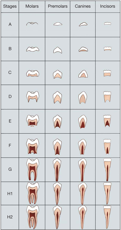
Fig. 1 Schematic illustration shows the modified Demirjian stages18 used to identify tooth development.

Fig. 2 Description and illustration reveals the eruption of permanent teeth by using Bengston's stages20 modified by AlQahtani et al.16 Position 1: When the occlusal or incisal surface is covered entirely by bone. Position 2: When the occlusal or incisal surface breaks through the crest of the alveolar bone. Position 3: When the occlusal or incisal surface is midway between alveolar bone and the occlusal plane. Position 4: Occlusal or incisal surface is in the occlusal plane.
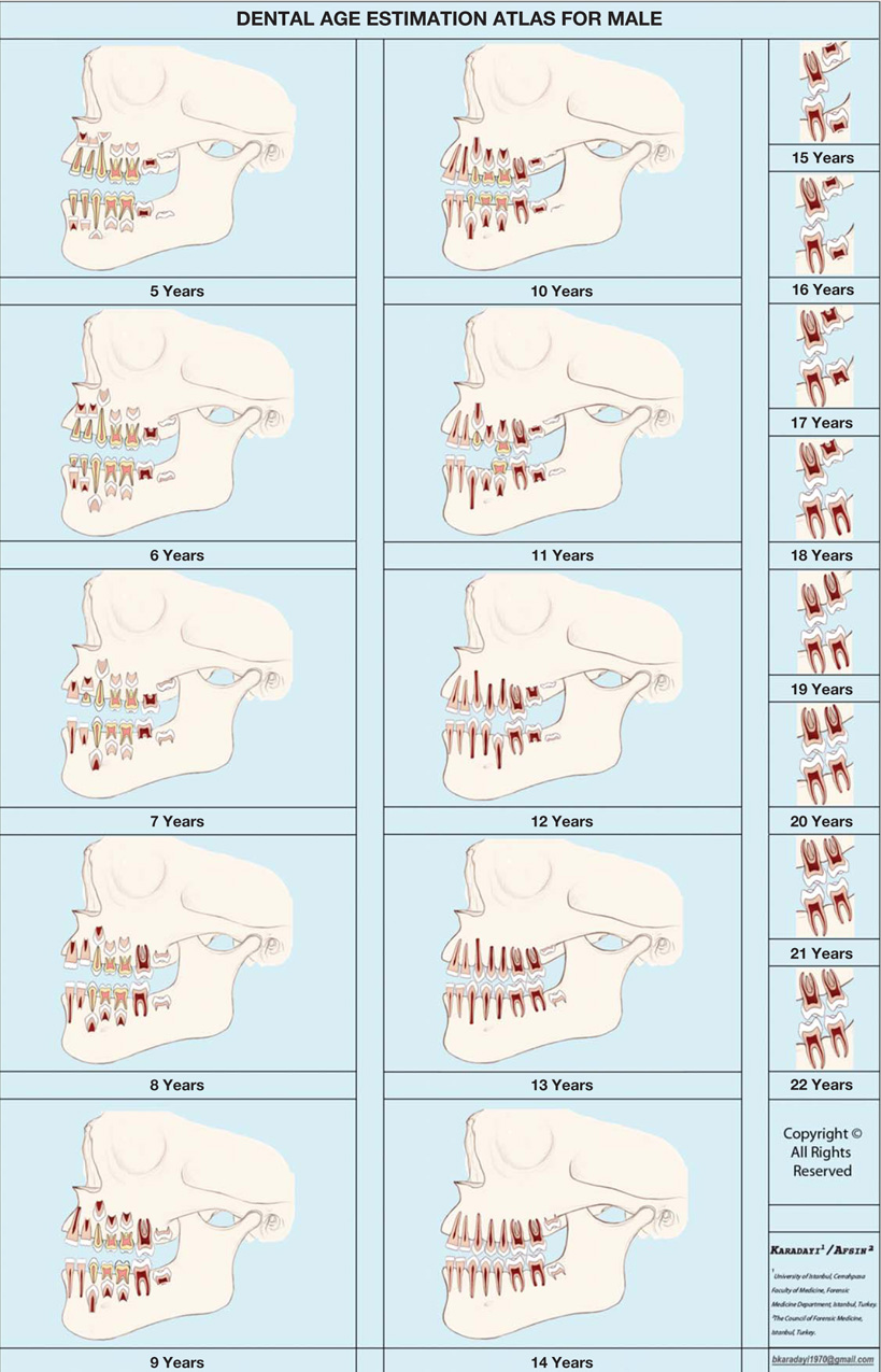
Fig. 3 Atlas of human tooth development and eruption for males. Age group: Group 5 includes all individuals aged 4.50-5.49 years; Group 6 includes all individuals aged 5.50-6.49 years etc.

Fig. 4 Atlas of human tooth development and eruption for females. Age group: Group 5 includes all individuals aged 4.50-5.49 years; Group 6 includes all individuals aged 5.50-6.49 years etc.
Reference
-
1. Introna F, Campobasso CP. Biological legal age of living individuals. In : Schmitt A, Cunha E, Pinheiro J, editors. Forensic anthropology medicine: complementary sciences from recovery to cause of death. Totowa, NJ: Humana Press;2006. p. 57–82.2. Demirjian A, Buschang PH, Tanguay R, Patterson DK. Interrelationships among measures of somatic, skeletal, dental, and sexual maturity. Am J Orthod. 1985; 88:433–438.
Article3. Cunha E, Baccino E, Martrille L, Ramsthaler F, Prieto J, Schuliar Y, et al. The problem of aging human remains and living individuals: a review. Forensic Sci Int. 2009; 193:1–13.
Article4. Choi JH, Kim CY. A study of correlation between the development of the third molar and second molar as an aid in age determination. J Korean Acad Oral Med. 1991; 16:121–136.5. Almeida MS, Pontual Ados A, Beltrão RT, Beltrão RV, Pontual ML. The chronology of second molar development in Brazilians and its application to forensic age estimation. Imaging Sci Dent. 2013; 43:1–6.
Article6. Tunc ES, Koyuturk AE. Dental age assessment using Demirjian's method on northern Turkish children. Forensic Sci Int. 2008; 175:23–26.
Article7. Kırzıoğlu Z, Ceyhan D. Accuracy of different dental age estimation methods on Turkish children. Forensic Sci Int. 2012; 216:61–67.
Article8. Phillips VM, van Wyk Kotze TJ. Testing standard methods of dental age estimation by Moorrees, Fanning and Hunt and Demirjian, Goldstein and Tanner on three South African children samples. J Forensic Odontostomatol. 2009; 27:20–28.9. Karataş OH, Öztürk F, Dedeoğlu N, Çolak C, Altun O. Dental age assessment: the applicability of Demirjian method in southwestern of eastern Anatolia region Turkish children. Cumhuriyet Dent J. 2012; 15:130–137.
Article10. Maber M, Liversidge HM, Hector MP. Accuracy of age estimation of radiographic methods using developing teeth. Forensic Sci Int. 2006; 159:Suppl 1. S68–S73.
Article11. Warren MW, Smith KR, Stubblefield PR, Martin SS, Walsh-Haney HA. Use of radiographic atlases in a mass fatality. J Forensic Sci. 2000; 45:467–470.
Article12. Logan WH, Kronfeld R. Development of the human jaws and surrounding structures from birth to the age of fifteen years. J Am Dent Assoc. 1933; 20:379–427.
Article13. Schour I, Massler M. The development of the human dentition. J Am Dent Assoc. 1941; 28:1153–1160.14. Ubelaker DH. Human skeletal remains. Chicago: Aldine;1978.15. Gustafson G, Koch G. Age estimation up to 16 years of age based on dental development. Odontol Revy. 1974; 25:297–306.16. AlQahtani SJ, Hector MP, Liversidge HM. Brief communication: the London atlas of human tooth development and eruption. Am J Phys Anthropol. 2010; 142:481–490.
Article17. Blenkin M, Taylor J. Age estimation charts for a modern Australian population. Forensic Sci Int. 2012; 221:106–112.
Article18. Demirjian A, Goldstein H, Tanner JM. A new system of dental age assessment. Hum Biol. 1973; 45:211–227.19. Moorrees CF, Fanning EA, Hunt EE Jr. Formation and resorption of three deciduous teeth in children. Am J Phys Anthropol. 1963; 21:205–213.
Article20. Bengston RG. A study of the time of eruption and root development of the permanent teeth between six and thirteen years. Dent Res Grad Study. 1935; 35:3–9.21. Landis JR, Koch GG. The measurement of observer agreement for categorical data. Biometrics. 1977; 33:159–174.
Article22. Estrela C, Neto JV, Bueno MR, Guedes OA, Porto OC, Pécora JD. Linear measurements of human permanent dental development stages using cone-beam computed tomography: a preliminary study. Dental Press J Orthod. 2010; 15:44–78.23. Makkad RS, Balani A, Chaturvedi SS, Tanwani T, Agrawal A, Hamdani S. Reliability of panoramic radiography in chronological age estimation. J Forensic Dent Sci. 2013; 5:129–133.
Article24. Alt KW, Rösing FW, Techler-Nicola M. Dental anthropology: fundamentals, limits, and prospects. Wien, New York: Springer Press;1998.25. Nolla CM. The development of the permanent teeth. J Dent Child. 1960; 27:254–266.26. Moorrees CF, Fanning EA, Hunt EE Jr. Age variation of formation stages for ten permanent teeth. J Dent Res. 1963; 42:1490–1502.
Article27. Haavikko K. The formation and the alveolar and clinical eruption of the permanent teeth. An orthopantomographic study. Suom Hammaslaak Toim. 1970; 66:103–170.28. Karadayi B, Kaya A, Kolusayın MO, Karadayi S, Afsin H, Ozaslan A. Radiological age estimation: based on third molar mineralization and eruption in Turkish children and young adults. Int J Legal Med. 2012; 126:933–942.
Article29. Zeng DL, Wu ZL, Cui MY. Chronological age estimation of third molar mineralization of Han in southern China. Int J Legal Med. 2010; 124:119–123.
Article30. Dhanjal KS, Bhardwaj MK, Liversidge HM. Reproducibility of radiographic stage assessment of third molars. Forensic Sci Int. 2006; 159:Suppl 1. S74–S77.
Article31. Harris EF. Mineralization of the mandibular third molar: a study of American blacks and whites. Am J Phys Anthropol. 2007; 132:98–109.
Article32. Thorson J, Hägg U. The accuracy and precision of the third mandibular molar as an indicator of chronological age. Swed Dent J. 1991; 15:15–22.33. Solari AC, Abramovitch K. The accuracy and precision of third molar development as an indicator of chronological age in Hispanics. J Forensic Sci. 2002; 47:531–535.
Article34. Butti AC, Clivio A, Ferraroni M, Spada E, Testa A, Salvato A. Häävikko's method to assess dental age in Italian children. Eur J Orthod. 2009; 31:150–155.35. Ogodescu AE, Bratu E, Tudor A, Ogodescu A. Estimation of child's biological age based on tooth development. Rom J Leg Med. 2011; 19:115–124.
Article36. Bolaños MV, Moussa H, Manrique MC, Bolaños MJ. Radiographic evaluation of third molar development in Spanish children and young people. Forensic Sci Int. 2003; 133:212–219.
Article37. Willershausen B, Löffler N, Schulze R. Analysis of 1202 orthopantograms to evaluate the potential of forensic age determination based on third molar developmental stages. Eur J Med Res. 2001; 28:377–384.38. Cantekin K, Yilmaz Y, Demirci T, Celikoglu M. Morphologic analysis of third-molar mineralization for eastern Turkish children and youth. J Forensic Sci. 2012; 57:531–534.
Article39. Sisman Y, Uysal T, Yagmur F, Ramoglu SI. Third-molar development in relation to chronologic age in Turkish children and young adults. Angle Orthod. 2007; 77:1040–1045.
Article40. Mincer HH, Harris EF, Berryman HE. The A.B.F.O. study of third molar development and its use as an estimator of chronological age. J Forensic Sci. 1993; 38:379–390.
Article41. Kullman L. Accuracy of two dental and one skeletal age estimation method in Swedish adolescents. Forensic Sci Int. 1995; 75:225–236.
Article42. Schmeling A, Olze A, Pynn BR, Kraul V, Schulz R, Heinecke A, et al. Dental age estimation based on third molar eruption in First Nation people of Canada. J Forensic Odontostomatol. 2010; 28:32–38.43. Olze A, Peschke C, Schulz R, Schmeling A. Studies of the chronological course of wisdom tooth eruption in a German population. J Forensic Leg Med. 2008; 15:426–429.
Article44. Hillson S. Teeth. 2nd ed. Cambridge: Cambridge University Press;2005.
- Full Text Links
-
- Actions
-
Cited
- CITED
-
- Close
- Share
-
- Similar articles
-
- The timing of tooth eruption and root development of permanent canine and premolars in Korean children
- Impacts of radicular development of lower third molars on its impaction
- The financial estimate of dental implant treatment about the National Health Insurance coverage for the Korean young adults
- A Case of Ectopic Tooth in the Maxillary Sinus Associated with a Mucocele
- Tooth Agenesis and Delay in Patients with Agenesis of Mandibular Second Premolars
