Korean J Ophthalmol.
2013 Jun;27(3):194-198. 10.3341/kjo.2013.27.3.194.
A Novel Computerized Visual Acuity Test for Children
- Affiliations
-
- 1Department of Ophthalmology, Hallym University College of Medicine, Chuncheon, Korea.
- 2Department of Biomechanics, Seoul National University College of Medicine, Seoul, Korea.
- 3Department of Ophthalmology, Seoul National University Bundang Hospital, Seoul National University College of Medicine, Seongnam, Korea. hjm@snu.ac.kr
- KMID: 1798057
- DOI: http://doi.org/10.3341/kjo.2013.27.3.194
Abstract
- PURPOSE
To investigate the efficacy of a computerized visual acuity test, the SNU visual acuity test for children.
METHODS
Fifty-six children, ranging from 1 to 5 years of age, were included. In a dark room, children gazed at and followed a circular dot with 50% contrast moving at a fixed velocity of 10 pixels/sec on a computer monitor. Eye movement was captured using a charge coupled device camera and was expressed as coordinates on a graph. Movements of the eye and dot were superimposed on a graph and analyzed. Minimum visualized dot diameters were compared to the Teller visual acuity.
RESULTS
Ten eyes (8.9%) of six children failed to perform the Teller visual acuity test, and two eyes (1.8%) of one patient failed to perform the SNU visual acuity test. The observed Teller visual acuity and SNU visual acuity were significantly correlated (p < 0.001). Visual angle degrees converted from the Teller visual acuity and SNU visual acuity were also significantly correlated (p < 0.001).
CONCLUSION
The SNU visual acuity using moving targets correlated well with Teller visual acuity and was more applicable than the Teller acuity test. Therefore, the SNU visual acuity test has potential clinical applications for children.
Keyword
MeSH Terms
Figure
-

Fig. 1 The SNU visual acuity test. The test equipment includes two monitors, one for an examiner (A) and another for the examinee (B). (A) Top left: tracking marker on the testing headband. Pupils can be observed via camera capture. This figure shows the volunteer's face for development of this test. Top right: the target on the monitor moved according to the examiner's manipulation of the mouse. Bottom: eye and target movement were superimposed and recorded on a graph. (B) The target moves at a fixed speed on a monitor with a black background.
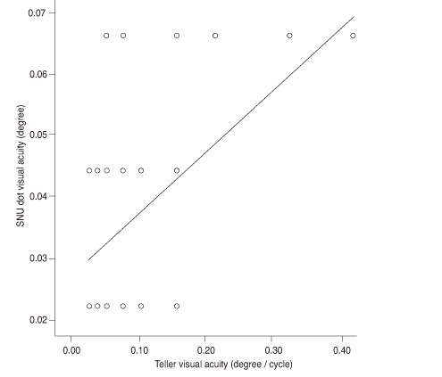
Fig. 2 Correlation between Teller visual acuity angle and SNU child visual acuity angle (p < 0.001, Pearson correlation test).
Reference
-
1. Lamkin JC. Can this baby see? Estimation of visual acuity in the preverbal child. Int Ophthalmol Clin. 1992. 32:1–23.2. Dobson V, Quinn GE, Biglan AW, et al. Acuity card assessment of visual function in the cryotherapy for retinopathy of prematurity trial. Invest Ophthalmol Vis Sci. 1990. 31:1702–1708.3. McDonald MA. Assessment of visual acuity in toddlers. Surv Ophthalmol. 1986. 31:189–210.4. Dobson V, Carpenter NA, Bonvalot K, Bossler J. The acuity card procedure: interobserver agreement in infants with perinatal complications. Clin Vis Sci. 1990. 6:39–48.5. Dobson V, Luna B. Prototype and Teller acuity cards yield similar acuities in infants and young children despite stimulus differences. Clin Vis Sci. 1993. 8:395–400.6. Hutchinson TE, White KP Jr, Martin WN, et al. Human-computer interaction using eye-gaze input. IEEE Trans Syst Man Cybern. 1989. 19:1527–1534.7. Riggs LA. Graham CH, editor. Visual acuity. Vision and visual perception. 1965. New York: John Wiley & Sons;321–349.8. Kushner BJ, Lucchese NJ, Morton GV. Grating visual acuity with Teller cards compared with Snellen visual acuity in literate patients. Arch Ophthalmol. 1995. 113:485–493.9. Hoyt CS. Cryotherapy for retinopathy of prematurity: 3 1/2-year outcome for both structure and function. Arch Ophthalmol. 1993. 111:319–320.10. Spierer A, Royzman Z, Chetrit A, et al. Vision screening of preverbal children with Teller acuity cards. Ophthalmology. 1999. 106:849–854.11. Park MG, Kim JW, Tchah HW, et al. Sensitivity, specificity and efficiency of Teller acuity cards for detecting amblyopia. J Korean Ophthalmol Soc. 1990. 31:697–701.12. Graham CH. Graham CH, editor. Perception of movement. Vision and visual perception. 1965. New York: John Wiley & Sons;575–588.13. Ho CS, Paul PS, Asirvatham A, et al. Abnormal spatial selection and tracking in children with amblyopia. Vision Res. 2006. 46:3274–3283.14. Kiorpes L, Tang C, Movshon JA. Sensitivity to visual motion in amblyopic macaque monkeys. Vis Neurosci. 2006. 23:247–256.15. Kirschen DG, Rosenbaum AL, Ballard EA. The dot visual acuity test: a new acuity test for children. J Am Optom Assoc. 1983. 54:1055–1059.16. Vuong QC, Hof AF, Bulthoff HH, Thornton IM. An advantage for detecting dynamic targets in natural scenes. J Vis. 2006. 6:87–96.17. Thornton IM, Kourtzi Z. A matching advantage for dynamic human faces. Perception. 2002. 31:113–132.
- Full Text Links
-
- Actions
-
Cited
- CITED
-
- Close
- Share
-
- Similar articles
-
- The Clinical Usefulness of the Teller Acuity Cards Test in Preverbal Children
- Relationship between Uncorrected Visual Acuity and Refractive Error in Visual Acuity Test of Children
- Visual Acuity Charts Comparison for Preschool Vision Screening
- Change of Visual Acuity and Astigmatism after Operation in Epiblepharon Children
- The Clinical Interpretation of Teller Acuity Card Test in the Diagnosis of Amblyopia
