Comparison of Three-Dimensional Isotropic and Two-Dimensional Conventional Indirect MR Arthrography for the Diagnosis of Rotator Cuff Tears
- Affiliations
-
- 1Department of Radiology, Samsung Medical Center, Sungkyunkwan University School of Medicine, Seoul 135-710, Korea. youngcheol.yoon@gmail.com
- 2Joeun Madi Hospital, Seoul 130-032, Korea.
- 3Department of Radiology, Soonchunhyang University Bucheon Hospital, Bucheon 420-767, Korea.
- 4Department of Orthopedic Surgery, Samsung Medical Center, Sungkyunkwan University School of Medicine, Seoul 135-710, Korea.
- KMID: 1794649
- DOI: http://doi.org/10.3348/kjr.2014.15.6.771
Abstract
OBJECTIVE
To compare the accuracy between a three-dimensional (3D) indirect isotropic T1-weighted fast spin-echo (FSE) magnetic resonance (MR) arthrography and a conventional two-dimensional (2D) T1-weighted sequences of indirect MR arthrography for diagnosing rotator cuff tears.
MATERIALS AND METHODS
The study was approved by our Institutional Review Board. In total, 205 patients who had undergone indirect shoulder MR arthrography followed by arthroscopic surgery for 206 shoulders were included in this study. Both conventional 2D T1-weighted FSE sequences and 3D isotropic T1-weighted FSE sequence were performed in all patients. Two radiologists evaluated the images for the presence of full- or partial-thickness tears in the supraspinatus-infraspinatus (SSP-ISP) tendons and tears in the subscapularis (SSC) tendons. Using the arthroscopic findings as the reference standard, the diagnostic performances of both methods were analyzed by the area under the receiver operating characteristic curve (AUC).
RESULTS
Arthroscopy confirmed 165 SSP-ISP tendon tears and 103 SSC tendon tears. For diagnosing SSP-ISP tendon tears, the AUC values were 0.964 and 0.989 for the 2D sequences and 3D T1-weighted FSE sequence, respectively, in reader I and 0.947 and 0.963, respectively, in reader II. The AUC values for diagnosing SSC tendon tears were 0.921 and 0.925, respectively, for reader I and 0.856 and 0.860, respectively, for reader II. There was no significant difference between the AUC values of the 2D and 3D sequences in either reader for either type of tear.
CONCLUSION
3D indirect isotropic MR arthrography with FSE sequence and the conventional 2D arthrography are not significantly different in terms of accuracy for diagnosing rotator cuff tears.
MeSH Terms
-
Adolescent
Adult
Aged
Area Under Curve
Female
Humans
Imaging, Three-Dimensional
*Magnetic Resonance Imaging
Male
Middle Aged
ROC Curve
Retrospective Studies
Rotator Cuff/injuries/pathology/*radiography
Sensitivity and Specificity
Shoulder Joint/injuries/pathology/*radiography
Tendons/pathology/radiography
Young Adult
Figure
-
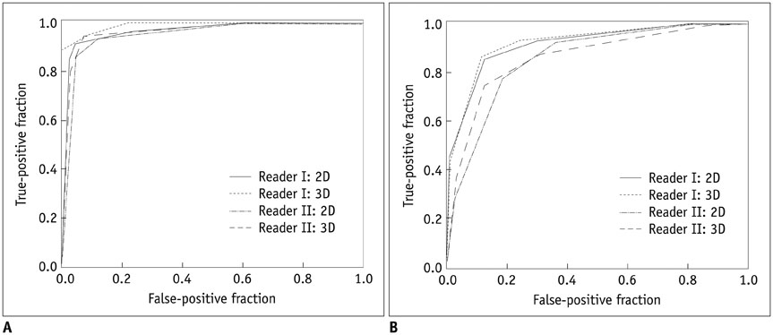
Fig. 1 Graphs showing receiver operating characteristic curves of readers I and II for detection of supraspinatus-infraspinatus (A) and subscapularis tendon tears (B) using two-dimensional (2D) and three-dimensional (3D) sequences.
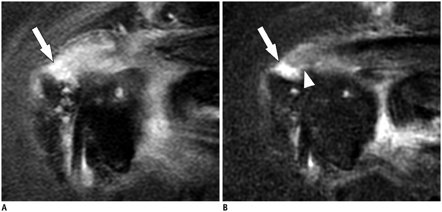
Fig. 2 Arthroscopically proven full-thickness tear of supraspinatus (SSP) tendon of right shoulder in 73-year-old woman. Fat-suppressed T1-weighted oblique coronal image (A) shows tendinous discontinuity of SSP tendon anterior portion at bursal side (arrow), but extension to articular side was not definite. Three-dimensional (3D) isotropic fat-suppressed T1-weighted image in arbitrary oblique coronal plane (B) demonstrates full-thickness tear of SSP tendon with retraction of musculotendinous junction, exposing humeral head (arrow). Small gap filled with contrast media is suspected at articular side of SSP tendon (arrowhead). It was correctly diagnosed only by using 3D isotropic sequence by both readers.
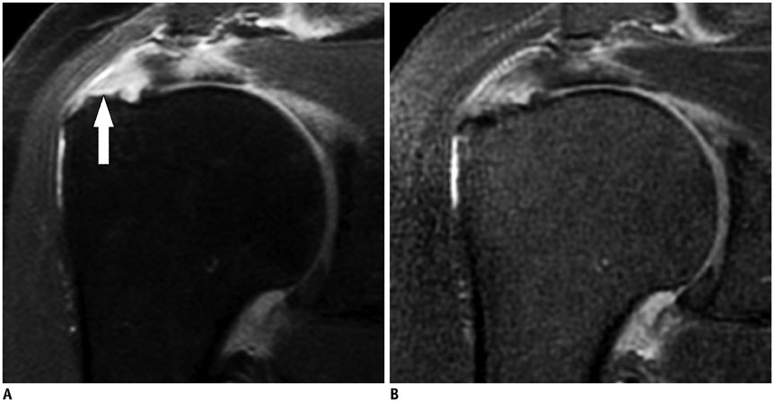
Fig. 3 Arthroscopically proven bursal-sided partial-thickness tear of supraspinatus (SSP) tendon of right shoulder in 49-year-old woman. Fat-suppressed T1-weighted oblique coronal image (A) shows focal defect filled with high signal at SSP tendon bursal side (arrow), which was not obvious on three-dimensional isotropic fat-suppressed T1-weighted oblique coronal image (B). It was correctly diagnosed only by using two-dimensional sequence by both readers.
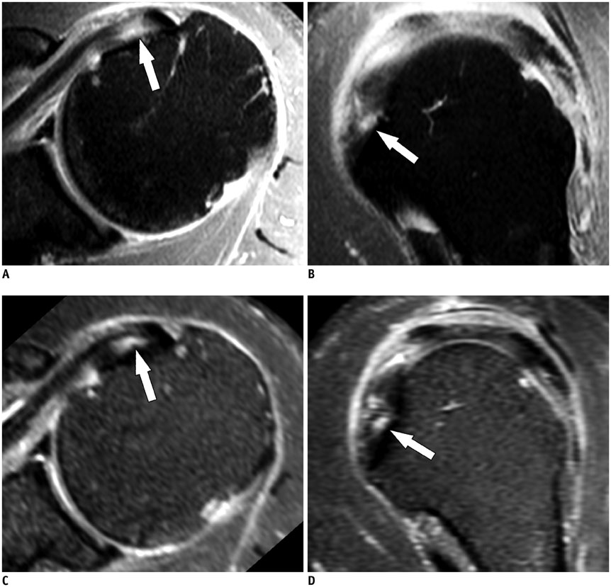
Fig. 4 Arthroscopically proven tear of subscapularis (SSC) tendon of left shoulder in 59-year-old man. Fat-suppressed T1-weighted axial (A) oblique sagittal (B) images and three-dimensional isotropic fat-suppressed T1-weighted axial (C), oblique sagittal (D) reformatted images show focal high signal intensity in articular side of SSC tendon (arrows), which was correctly interpreted as tear by both readers using both sequences.
Cited by 1 articles
-
Diagnosis of Rotator Cuff Tears with Non-Arthrographic MR Imaging: 3D Fat-Suppressed Isotropic Intermediate-Weighted Turbo Spin-Echo Sequence versus Conventional 2D Sequences at 3T
Won Sun Hong, Won-Hee Jee, So-Yeon Lee, Chang-Woo Chun, Joon-Yong Jung, Yang-Soo Kim
Investig Magn Reson Imaging. 2018;22(4):229-239. doi: 10.13104/imri.2018.22.4.229.
Reference
-
1. Hodler J, Kursunoglu-Brahme S, Snyder SJ, Cervilla V, Karzel RP, Schweitzer ME, et al. Rotator cuff disease: assessment with MR arthrography versus standard MR imaging in 36 patients with arthroscopic confirmation. Radiology. 1992; 182:431–436.2. Massengill AD, Seeger LL, Yao L, Gentili A, Shnier RC, Shapiro MS, et al. Labrocapsular ligamentous complex of the shoulder: normal anatomy, anatomic variation, and pitfalls of MR imaging and MR arthrography. Radiographics. 1994; 14:1211–1223.3. Palmer WE, Caslowitz PL, Chew FS. MR arthrography of the shoulder: normal intraarticular structures and common abnormalities. AJR Am J Roentgenol. 1995; 164:141–146.4. Sahin G, Demirtasş M. An overview of MR arthrography with emphasis on the current technique and applicational hints and tips. Eur J Radiol. 2006; 58:416–430.5. Uri DS, Kneeland JB, Dalinka MK. Update in shoulder magnetic resonance imaging. Magn Reson Q. 1995; 11:21–44.6. Bergin D, Schweitzer ME. Indirect magnetic resonance arthrography. Skeletal Radiol. 2003; 32:551–558.7. Dinauer PA, Flemming DJ, Murphy KP, Doukas WC. Diagnosis of superior labral lesions: comparison of noncontrast MRI with indirect MR arthrography in unexercised shoulders. Skeletal Radiol. 2007; 36:195–202.8. Lee MJ, Motamedi K, Chow K, Seeger LL. Gradient-recalled echo sequences in direct shoulder MR arthrography for evaluating the labrum. Skeletal Radiol. 2008; 37:19–25.9. Vahlensieck M, Sommer T, Textor J, Pauleit D, Lang P, Genant HK, et al. Indirect MR arthrography: techniques and applications. Eur Radiol. 1998; 8:232–235.10. Yagci B, Manisali M, Yilmaz E, Ozkan M, Ekin A, Ozaksoy D, et al. Indirect MR arthrography of the shoulder in detection of rotator cuff ruptures. Eur Radiol. 2001; 11:258–262.11. Jung JY, Yoon YC, Yi SK, Yoo J, Choe BK. Comparison study of indirect MR arthrography and direct MR arthrography of the shoulder. Skeletal Radiol. 2009; 38:659–667.12. Kijowski R, Davis KW, Woods MA, Lindstrom MJ, De Smet AA, Gold GE, et al. Knee joint: comprehensive assessment with 3D isotropic resolution fast spin-echo MR imaging--diagnostic performance compared with that of conventional MR imaging at 3.0 T. Radiology. 2009; 252:486–495.13. Kwon JW, Yoon YC, Choi SH. Three-dimensional isotropic T2-weighted cervical MRI at 3T: comparison with two-dimensional T2-weighted sequences. Clin Radiol. 2012; 67:106–113.14. Stevens KJ, Wallace CG, Chen W, Rosenberg JK, Gold GE. Imaging of the wrist at 1.5 Tesla using isotropic three-dimensional fast spin echo cube. J Magn Reson Imaging. 2011; 33:908–915.15. Subhas N, Kao A, Freire M, Polster JM, Obuchowski NA, Winalski CS. MRI of the knee ligaments and menisci: comparison of isotropic-resolution 3D and conventional 2D fast spin-echo sequences at 3 T. AJR Am J Roentgenol. 2011; 197:442–450.16. Jung JY, Yoon YC, Kwon JW, Ahn JH, Choe BK. Diagnosis of internal derangement of the knee at 3.0-T MR imaging: 3D isotropic intermediate-weighted versus 2D sequences. Radiology. 2009; 253:780–787.17. Gold GE, Busse RF, Beehler C, Han E, Brau AC, Beatty PJ, et al. Isotropic MRI of the knee with 3D fast spin-echo extended echo-train acquisition (XETA): initial experience. AJR Am J Roentgenol. 2007; 188:1287–1293.18. Notohamiprodjo M, Horng A, Pietschmann MF, Müller PE, Horger W, Park J, et al. MRI of the knee at 3T: first clinical results with an isotropic PDfs-weighted 3D-TSE-sequence. Invest Radiol. 2009; 44:585–597.19. Choo HJ, Lee SJ, Kim OH, Seo SS, Kim JH. Comparison of three-dimensional isotropic T1-weighted fast spin-echo MR arthrography with two-dimensional MR arthrography of the shoulder. Radiology. 2012; 262:921–931.20. Jung JY, Jee WH, Park MY, Lee SY, Kim YS. Supraspinatus tendon tears at 3.0 T shoulder MR arthrography: diagnosis with 3D isotropic turbo spin-echo SPACE sequence versus 2D conventional sequences. Skeletal Radiol. 2012; 41:1401–1410.21. Minagawa H, Itoi E, Konno N, Kido T, Sano A, Urayama M, et al. Humeral attachment of the supraspinatus and infraspinatus tendons: an anatomic study. Arthroscopy. 1998; 14:302–306.22. Mochizuki T, Sugaya H, Uomizu M, Maeda K, Matsuki K, Sekiya I, et al. Humeral insertion of the supraspinatus and infraspinatus. New anatomical findings regarding the footprint of the rotator cuff. J Bone Joint Surg Am. 2008; 90:962–969.23. Clark JM, Harryman DT 2nd. Tendons, ligaments, and capsule of the rotator cuff. Gross and microscopic anatomy. J Bone Joint Surg Am. 1992; 74:713–725.24. Oh DK, Yoon YC, Kwon JW, Choi SH, Jung JY, Bae S, et al. Comparison of indirect isotropic MR arthrography and conventional MR arthrography of labral lesions and rotator cuff tears: a prospective study. AJR Am J Roentgenol. 2009; 192:473–479.25. Opsha O, Malik A, Baltazar R, Primakov D, Beltran S, Miller TT, et al. MRI of the rotator cuff and internal derangement. Eur J Radiol. 2008; 68:36–56.26. Pfirrmann CW, Zanetti M, Weishaupt D, Gerber C, Hodler J. Subscapularis tendon tears: detection and grading at MR arthrography. Radiology. 1999; 213:709–714.27. DeLong ER, DeLong DM, Clarke-Pearson DL. Comparing the areas under two or more correlated receiver operating characteristic curves: a nonparametric approach. Biometrics. 1988; 44:837–845.28. Hawass NE. Comparing the sensitivities and specificities of two diagnostic procedures performed on the same group of patients. Br J Radiol. 1997; 70:360–366.29. Kijowski R, Gold GE. Routine 3D magnetic resonance imaging of joints. J Magn Reson Imaging. 2011; 33:758–771.30. Hill NB Jr, Bucchieri JS, Shon F, Miller TT, Rosenwasser MP. Magnetic resonance imaging of injury to the medial collateral ligament of the elbow: a cadaver model. J Shoulder Elbow Surg. 2000; 9:418–422.31. Jung JY, Yoon YC, Choi SH, Kwon JW, Yoo J, Choe BK. Three-dimensional isotropic shoulder MR arthrography: comparison with two-dimensional MR arthrography for the diagnosis of labral lesions at 3.0 T. Radiology. 2009; 250:498–505.32. Magee T. Can isotropic fast gradient echo imaging be substituted for conventional T1 weighted sequences in shoulder MR arthrography at 3 Tesla? J Magn Reson Imaging. 2007; 26:118–122.33. Stevens KJ, Busse RF, Han E, Brau AC, Beatty PJ, Beaulieu CF, et al. Ankle: isotropic MR imaging with 3D-FSE-cube--initial experience in healthy volunteers. Radiology. 2008; 249:1026–1033.34. Yao L, Pitts JT, Thomasson D. Isotropic 3D fast spin-echo with proton-density-like contrast: a comprehensive approach to musculoskeletal MRI. AJR Am J Roentgenol. 2007; 188:W199–W201.35. Ristow O, Stehling C, Krug R, Steinbach L, Sabo G, Ambekar A, et al. Isotropic 3-dimensional fast spin echo imaging versus standard 2-dimensional imaging at 3.0 T of the knee: artificial cartilage and meniscal lesions in a porcine model. J Comput Assist Tomogr. 2010; 34:260–269.36. Seo JM, Yoon YC, Kwon JW. 3D isotropic turbo spin-echo intermediate-weighted sequence with refocusing control in knee imaging: comparison study with 3D isotropic fast-field echo sequence. Acta Radiol. 2011; 52:1119–1124.37. Rybak LD, La Rocca Vieira R, Recht M, Shepard T, Wiggins G, Babb J, et al. Preliminary study of 1.5-T MR arthrography of the shoulder with 3D isotropic intermediate-weighted turbo spin echo. AJR Am J Roentgenol. 2012; 199:W107–W113.38. Jung JY, Jee WH, Park MY, Lee SY, Kim YS. SLAP tears: diagnosis using 3-T shoulder MR arthrography with the 3D isotropic turbo spin-echo space sequence versus conventional 2D sequences. Eur Radiol. 2013; 23:487–495.39. Johnson G, Wadghiri YZ, Turnbull DH. 2D multislice and 3D MRI sequences are often equally sensitive. Magn Reson Med. 1999; 41:824–828.40. Herold T, Bachthaler M, Hamer OW, Hente R, Feuerbach S, Fellner C, et al. Indirect MR arthrography of the shoulder: use of abduction and external rotation to detect full- and partial-thickness tears of the supraspinatus tendon. Radiology. 2006; 240:152–160.41. Van Dyck P, Gielen JL, Veryser J, Weyler J, Vanhoenacker FM, Van Glabbeek F, et al. Tears of the supraspinatus tendon: assessment with indirect magnetic resonance arthrography in 67 patients with arthroscopic correlation. Acta Radiol. 2009; 50:1057–1063.42. Yoo JC, Jun BJ, Shin SJ, Mcgarry MH, Lee TQ. Subscapularis footprint anatomy revisited with 3-dimensional perspective and its relationship with supraspinatus 1st facet. Arthroscopy. 2013; 29:e95.
- Full Text Links
-
- Actions
-
Cited
- CITED
-
- Close
- Share
-
- Similar articles
-
- Comparative Study of MR-arthrography and Arthroscopy in Partial Thickness Rotator Cuff Tears
- Feasibility of Ultrasonography and MR Arthrography during Evaluation of Rotator Cuff Injury
- Comparisons of the Various Partial-Thickness Rotator Cuff Tears on MR Arthrography and Arthroscopic Correlation
- Arthrographic Pitfalls in the Diagnosis of Full-Thickness Tears of the Rotator Cuff: A Case Report
- Characteristics of Magnetic Resonance Arthrography Findings in Traumatic Posterosuperior Rotator Cuff Tears
