Extraskeletal Mesenchymal Chondrosarcoma of the Carotid Space: A Case Report
- Affiliations
-
- 1Department of Radiology, Bucheon Hospital, Soonchunhyang University College of Medicine, Bucheon, Korea. hshong@schmc.ac.kr
- 2Department of Pathology, Bucheon Hospital, Soonchunhyang University College of Medicine, Bucheon, Korea.
- KMID: 1793888
- DOI: http://doi.org/10.3348/jksr.2015.72.5.324
Abstract
- Chondrosarcoma is a commonly encountered malignant cartilaginous tumor. However, only 1% of chondrosarcomas arise in the extraskeletal region. The pathologic types of this tumor include mesenchymal, myxoid, and low grade. A mesenchymal chondrosarcoma is a rare, highly malignant cartilaginous tumor that is rarely encountered, and it shows similar imaging features to other malignant soft-tissue tumors. Here, we report a mesenchymal chondrosarcoma presenting as a palpable mass in the neck, arising in the carotid space, which is also known as the retrostyloid parapharyngeal space.
Figure
-

Fig. 1 A 64-year-old man presented with a gradually increasing, palpable neck mass and 11 kg weight loss in 9 months. A. Precontrast CT reveals soft tissue density mass in the left carotid space. Dense calcifications with a ring-and-arc pattern representing chondroid calcifications is noted within the mass. B. Contrast enhanced CT reveals heterogeneous enhancing solid mass encasing the left common, internal, and external carotid arteries with splaying internal and external carotid arteries.
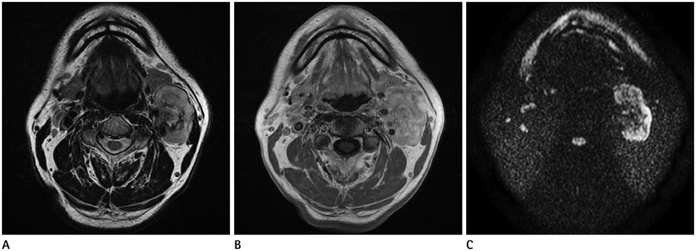
Fig. 2 The MR images of 64-year-old man with extraskeletal mesenchymal chondrosarcoma of the carotid space. A. The lesion shows heterogeneously hyperintense with heterogeneously low-signal foci representing a chondroid matrix on T2-weighted imaging and reveals invasion of surrounding structures with extension to the lowest crotch of the carotid bifurcation and encasement of the left external and internal carotid arteries. B. Contrast enhanced axial MRI scan shows well enhancing heterogeneous mass with low signal intensity foci in the left carotid space with splaying internal carotid artery and external carotid artery. The lesion shows isosignal intensity on T1-weighted imaging with low-signal-intensity foci (not shown). C. On diffusion-weighted imaging, this mass shows heterogeneous high-signal intensity and diffusion restriction on apparent diffusion coefficient mapping (not shown), suggestive of a malignant character of the tumor.
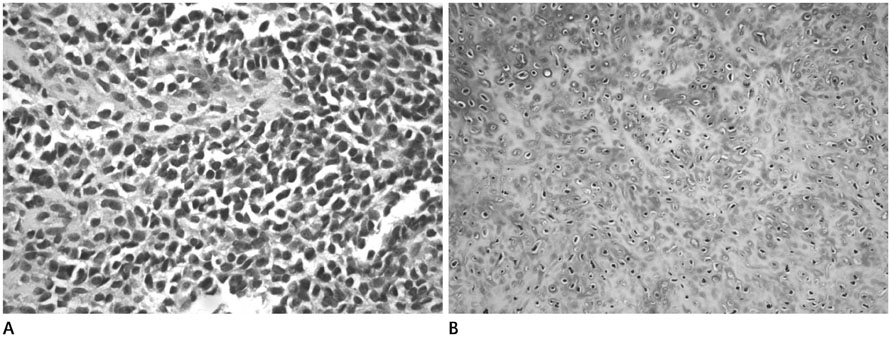
Fig. 3 Histopathological findings have a characteristic bimophic structure. A. One core reveals sheets of hypercellular, undifferentiated tumor cells. The other core shows a well-defined nodule of well-differentiated cartilage. A undifferentiated tumor cells have ovoid or elongated hyperchromatic nuclei and scanty, poorly outlined cytoplasm (hematoxylin-eosin, × 200). B. Chondroid component show well differentiated cartilage (hematoxylin-eosin, × 100).
Reference
-
1. Shapeero LG, Vanel D, Couanet D, Contesso G, Ackerman LV. Extraskeletal mesenchymal chondrosarcoma. Radiology. 1993; 186:819–826.2. Lightenstein L, Bernstein D. Unusual benign and malignant chondroid tumors of bone. A survey of some mesenchymal cartilage tumors and malignant chondroblastic tumors, including a few multicentric ones, as well as many atypical benign chondroblastomas and chondromyxoid fibromas. Cancer. 1959; 12:1142–1157.3. Nakashima Y, Unni KK, Shives TC, Swee RG, Dahlin DC. Mesenchymal chondrosarcoma of bone and soft tissue. A review of 111 cases. Cancer. 1986; 57:2444–2453.4. Bahr AL, Gayler BW. Cranial chondrosarcomas. Report of four cases and review of the literature. Radiology. 1977; 124:151–156.5. Mafee MF, Valbasson GE, Becker M. Imaging of the Head and Neck. Stuttgart: Georg Thieme Verlag;2012. p. 602–608.6. Som PM, Curtin HD. Head and Neck Imaging. Boston: Elsevier Health Sciences;2011. p. 1749–1779.7. Murphey MD, Walker EA, Wilson AJ, Kransdorf MJ, Temple HT, Gannon FH. From the archives of the AFIP: imaging of primary chondrosarcoma: radiologic-pathologic correlation. Radiographics. 2003; 23:1245–1278.8. Hashimoto N, Ueda T, Joyama S, Araki N, Beppu Y, Tatezaki S, et al. Extraskeletal mesenchymal chondrosarcoma: an imaging review of ten new patients. Skeletal Radiol. 2005; 34:785–792.9. Huvos AG, Rosen G, Dabska M, Marcove RC. Mesenchymal chondrosarcoma. A clinicopathologic analysis of 35 patients with emphasis on treatment. Cancer. 1983; 51:1230–1237.10. Banks KP, Ly JQ, Thompson LD, Michaelson PG, Davis SW. Mesenchymal chondrosarcoma of sinonasal cavity: a case report and brief literature review. Eur J Radiol Extra. 2004; 49:47–51.
- Full Text Links
-
- Actions
-
Cited
- CITED
-
- Close
- Share
-
- Similar articles
-
- A Case of Postoperative Chemotherapy of Extraskeletal Mesenchymal Chondrosarcoma
- Extraskeletal Mesenchymal Chondrosarcoma of the Mediastinum: A Case Report
- A Case Report of Extraskeletal Chondrosarcoma
- Fine Needle Aspiration Cytology of Extraskeletal Mesenchymal Chondrosarcoma
- A Case of Mesenchymal Chondrosarcoma In Pancreas
