Collision of Three Histologically Distinct Endometrial Cancers of the Uterus
- Affiliations
-
- 1Department of Pathology, College of Medicine, Hanyang University, Seoul, Korea.
- 2Department of Obstetrics and Gynecology, College of Medicine, Hanyang University, Seoul, Korea. chosh@hanyang.ac.kr
- KMID: 1792985
- DOI: http://doi.org/10.3346/jkms.2012.27.1.89
Abstract
- A collision tumor is defined by the presence of two separate masses in one organ, which are pathologically distinct. We described a 70-yr-old patient who complained of abnormal vaginal bleeding with a collision tumor of the uterine corpus. The patient received total hysterectomy, bilateral salphingo-oophorectomy, bilateral pelvic-paraaortic lymphadenectomy, omentectomy, and intraperitoneal chemotherapy. The uterine corpus revealed three separate masses, which were located at the fundus, anterior and posterior wall. Each tumor revealed three pathologically different components, which were malignant mixed mullerian tumor, papillary serous carcinoma, and endometrioid adenocarcinoma. Among these components, only the papillary serous carcinoma component invaded the underlying myometrium and metastasized to the regional lymph node. Adjuvant chemotherapy and radiation therapy were performed. The patient is still alive and has been healthy for the last 8 yr. We have reviewed previously reported cases of collision tumors which have occurred in the uterine corpus.
Keyword
MeSH Terms
-
Aged
Aromatase Inhibitors/therapeutic use
Carcinoma, Endometrioid/drug therapy/*pathology/surgery
Chemotherapy, Adjuvant
Cystadenocarcinoma, Papillary/drug therapy/*pathology/surgery
Endometrial Neoplasms/drug therapy/*pathology/surgery
Female
Humans
Hysterectomy
Immunohistochemistry
Keratins/metabolism
Lymphatic Metastasis
Mixed Tumor, Mullerian/drug therapy/*pathology/surgery
Nitriles/therapeutic use
Triazoles/therapeutic use
Tumor Suppressor Protein p53/metabolism
Figure
-
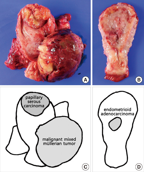
Fig. 1 Macroscopic appearance and schematic view of the hysterectomy specimen. (A) The opened uterus reveals a large polypoid mass in the anterior wall and a broad-based protruding mass in fundus. (B) The posterior wall of uterine corpus shows a slightly elevated mass. (C, D) Schematic view of three separate tumors with their pathologic diagnosis.
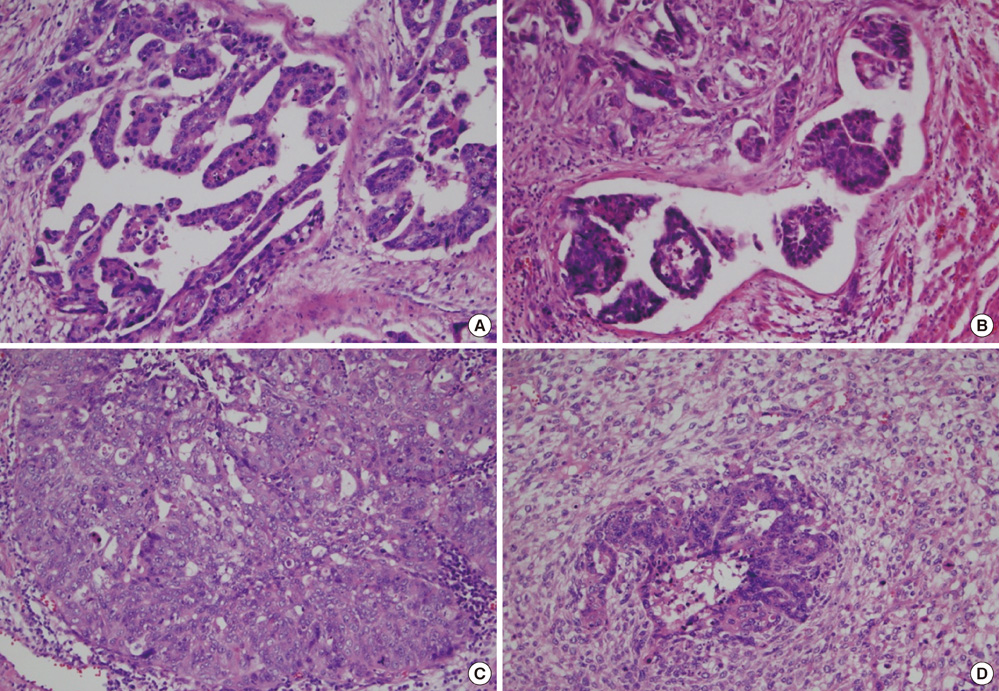
Fig. 2 Histopathology of the hysterectomy specimen HE stained. (A) Papillary serous carcinoma, which is found at fundus of uterine corpus, consists of pleomorphic tumor cells with papillary growth pattern. (B) The lymphovascular invasion is present at the periphery of the papillary serous carcinoma. (C) Section from posterior wall consists of endometrioid adenocarcinoma, showing glandular and solid growth pattern. (D) Section from polypoid mass reveals malignant mixed müllerian tumor, consisting of carcinomatous and sarcomatous components.
Reference
-
1. Van Eeden S, Nederlof PM, Taal BG, Offerhaus GJ, Van Velthuysen ML. A tumour with a neuroendocrine and papillary serous component: two or a pair? J Clin Pathol. 2002. 55:710–714.2. Lam KY, Khoo US, Cheung A. Collision of endometrioid carcinoma and stromal sarcoma of the uterus: a report of two cases. Int J Gynecol Pathol. 1999. 18:77–81.3. Gaertner EM, Farley JH, Taylor RR, Silver SA. Collision of uterine rhabdoid tumor and endometrioid adenocarcinoma: a case report and review of the literature. Int J Gynecol Pathol. 1999. 18:396–401.4. Lifschitz-Mercer B, Czernobilsky B, Dgani R, Dallenbach-Hellweg G, Moll R, Franke WW. Immunocytochemical study of an endometrial diffuse clear cell stromal sarcoma and other endometrial stromal sarcomas. Cancer. 1987. 59:1494–1499.5. Patwardhan JR, Gadgil RK. Collision tumour of the uterus. Indian J Cancer. 1969. 6:194–197.6. Shaco-Levy R, Manor E, Piura B, Ariel I. An unusual composite endometrial tumor combining papillary serous carcinoma and small cell carcinoma. Am J Surg Pathol. 2004. 28:1103–1106.7. Sreenan JJ, Hart WR. Carcinosarcomas of the female genital tract. A pathologic study of 29 metastatic tumors: further evidence for the dominant role of the epithelial component and the conversion theory of histogenesis. Am J Surg Pathol. 1995. 19:666–674.8. Takahashi Y, Inoue T. Hepatoid carcinoma of the uterus that collided with carcinosarcoma. Pathol Int. 2003. 53:323–326.9. Lewin K. Carcinoid tumors and the mixed (composite) glandular-endocrine cell carcinomas. Am J Surg Pathol. 1987. 11:71–86.10. Rose PG. Endometrial carcinoma. N Engl J Med. 1996. 335:640–649.
- Full Text Links
-
- Actions
-
Cited
- CITED
-
- Close
- Share
-
- Similar articles
-
- A Case of Endometrial Adenocarcinoma in a Double Uterus
- Collision Tumor of the Liver (Hepatocellular Carcinoma and Undifferentiated Sarcoma)
- Endometrial carcinoma arising in a bicornuate uterus
- A single horn endometrial carcinoma of a uterus bicornis unicollis
- A Case of Collision Tumor Composed of Dermatofibroma and Ito Nevus
