Autosomal Recessive Multiple Epiphyseal Dysplasia in a Korean Girl Caused by Novel Compound Heterozygous Mutations in the DTDST (SLC26A2) Gene
- Affiliations
-
- 1Department of Orthopaedic Surgery, Seoul National University Children's Hospital, Seoul, Korea. tjcho@snu.ac.kr
- 2Department of Radiology, Ajou University Hospital, Suwon, Korea.
- 3Department of Biochemistry and Molecular Medicine, Seoul National University College of Medicine, Seoul, Korea.
- 4Department of Laboratory Medicine, Seoul National University Children's Hospital, Seoul, Korea.
- KMID: 1792968
- DOI: http://doi.org/10.3346/jkms.2010.25.7.1105
Abstract
- Multiple epiphyseal dysplasia is caused by heterogenous genotypes involving more than six genes. Recessive mutations in the DTDST gene cause a phenotype of recessive multiple epiphyseal dysplasia (rMED). The authors report a 9-yr old Korean girl with the rMED phenotype having novel compound heterozygous mutations in the DTDST gene, which were inherited from both parents. This is the first Korean rMED case attributed to DTDST mutations, and expands the spectrum of diseases caused by DTDST mutations.
MeSH Terms
Figure
-
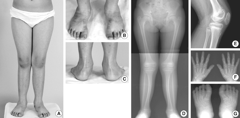
Fig. 1 The proband aged 9 yr. Mild genu valgum, forefoot adduction, and hindfoot valgus were noted (A-C). Radiographs showed genu valgum with flattening of epiphyses at the hips, knees, and ankles (D); double layered patella (E); shortening of the metacarpals and phalanges, premature physeal closure at the middle phalanges, cone-shaped epiphyses of the proximal phalanges (F); twisted, oblique arrays of the metatarsals, triangular medial cuneiforms (G).

Fig. 2 Sequencing results of the DTDST gene showed compound heterozygous mutations of c.485_486delTG and c.1153G>A in the proband. The c.485_486delTG mutation was inherited maternally, and the c.1153G>A point mutation paternally.
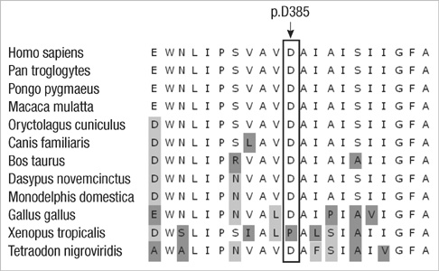
Fig. 3 Conservation of p.D385 in DTDST protein among different species.
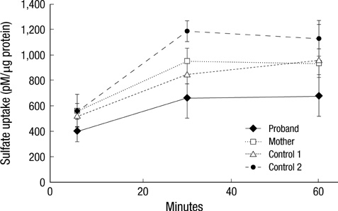
Fig. 4 Proband dermal fibroblast sulfate uptake was about 65-75% that of the normal control.
Reference
-
1. Jakkula E, Makitie O, Czarny-Ratajczak M, Jackson GC, Damignani R, Susic M, Briggs MD, Cole WG, Ala-Kokko L. Mutations in the known genes are not the major cause of MED; distinctive phenotypic entities among patients with no identified mutations. Eur J Hum Genet. 2005. 13:292–301.
Article2. Itoh T, Shirahama S, Nakashima E, Maeda K, Haga N, Kitoh H, Kosaki R, Ohashi H, Nishimura G, Ikegawa S. Comprehensive screening of multiple epiphyseal dysplasia mutations in Japanese population. Am J Med Genet A. 2006. 140:1280–1284.
Article3. Superti-Furga A, Neumann L, Riebel T, Eich G, Steinmann B, Spranger J, Kunze J. Recessively inherited multiple epiphyseal dysplasia with normal stature, club foot, and double layered patella caused by a DTDST mutation. J Med Genet. 1999. 36:621–624.4. Makitie O, Savarirayan R, Bonafe L, Robertson S, Susic M, Superti-Furga A, Cole WG. Autosomal recessive multiple epiphyseal dysplasia with homozygosity for C653S in the DTDST gene: double-layer patella as a reliable sign. Am J Med Genet A. 2003. 122:187–192.5. Ballhausen D, Bonafe L, Terhal P, Unger SL, Bellus G, Classen M, Hamel BC, Spranger J, Zabel B, Cohn DH, Cole WG, Hecht JT, Superti-Furga A. Recessive multiple epiphyseal dysplasia (rMED): phenotype delineation in eighteen homozygotes for DTDST mutation R279W. J Med Genet. 2003. 40:65–71.
Article6. Miyake A, Nishimura G, Futami T, Ohashi H, Chiba K, Toyama Y, Furuichi T, Ikegawa S. A compound heterozygote of novel and recurrent DTDST mutations results in a novel intermediate phenotype of Desbuquois dysplasia, diastrophic dysplasia, and recessive form of multiple epiphyseal dysplasia. J Hum Genet. 2008. 53:764–768.
Article7. Imauchi Y, Lombes M, Laine P, Sterkers O, Ferrary E, Grayeli AB. Glucocorticoids inhibit diastrophic dysplasia sulfate transporter activity in otosclerosis by interleukin-6. Laryngoscope. 2006. 116:1647–1650.
Article8. Hastbacka J, de la Chapelle A, Mahtani MM, Clines G, Reeve-Daly MP, Daly M, Hamilton BA, Kusumi K, Trivedi B, Weaver A, Coloma A, Lovett M, Buckler A, Kaitila I, Lander ES. The diastrophic dysplasia gene encodes a novel sulfate transporter: positional cloning by fine-structure linkage disequilibrium mapping. Cell. 1994. 78:1073–1087.
Article9. Rossi A, Bonaventure J, Delezoide AL, Cetta G, Superti-Furga A. Undersulfation of proteoglycans synthesized by chondrocytes from a patient with achondrogenesis type 1B homozygous for an L483P substitution in the diastrophic dysplasia sulfate transporter. J Biol Chem. 1996. 271:18456–18464.
Article10. Forlino A, Piazza R, Tiveron C, Della Torre S, Tatangelo L, Bonafe L, Gualeni B, Romano A, Pecora F, Superti-Furga A, Cetta G, Rossi A. A diastrophic dysplasia sulfate transporter (SLC26A2) mutant mouse: morphological and biochemical characterization of the resulting chondrodysplasia phenotype. Hum Molec Genet. 2005. 14:859–871.
Article11. Hastbacka J, Superti-Furga A, Wilcox WR, Rimoin DL, Cohn DH, Lander ES. Atelosteogenesis type II is caused by mutations in the diastrophic dysplasia sulfate-transporter gene (DTDST): evidence for a phenotypic series involving three chondrodysplasias. Am J Hum Genet. 1996. 58:255–262.12. Peterson HA. Skewfoot (forefoot adduction with heel valgus). J Pediatr Orthop. 1986. 6:24–30.
Article13. Ryoppy S, Poussa M, Merikanto J, Marttinen E, Kaitila I. Foot deformities in diastrophic dysplasia. An analysis of 102 patients. J Bone Joint Surg Br. 1992. 74:441–444.
Article
- Full Text Links
-
- Actions
-
Cited
- CITED
-
- Close
- Share
-
- Similar articles
-
- Novel Compound Heterozygous Mutations in CTSC Gene in a Chinese Family with Papillon–Lefevre Syndrome
- A compound heterozygous mutation in the FMO3 gene: the first pediatric case causes fish odor syndrome in Korea
- Novel Compound Heterozygous Mutations in the Vitamin D Receptor Gene in a Korean Girl with Hereditary Vitamin D Resistant Rickets
- Four Cases of Multiple Epiphyseal Dysplasia in One Family
- Two Novel Mutations in the Aquaporin 2 Gene in a Girl with Congenital Nephrogenic Diabetes Insipidus
