A Case of Rectal Mucosa-associated Lymphoid Tissue Lymphoma Diagnosed by Endoscopic Unroofing Technique
- Affiliations
-
- 1Division of Gastroenterology, Department of Internal Medicine, Gachon University Gil Medical Center, Incheon, Korea. junwonchung@hanmail.net
- 2Division of Hemato-oncology, Department of Internal Medicine, Gachon University Gil Medical Center, Incheon, Korea.
- 3Department of Pathology, Ewha Womans University School of Medicine, Seoul, Korea.
- KMID: 1792848
- DOI: http://doi.org/10.4166/kjg.2012.59.6.428
Abstract
- Mucosa-associated lymphoid tissue (MALT) lymphoma is a typical primary gastrointestinal lymphoma, particularly in the stomach. Although primary rectal lymphoma is rare, it may present as a subepithelial tumor. Several techniques have been proposed for a tissue diagnosis in subepithelial tumor, including endoscopic ultrasonography (EUS)-guided fine needle aspiration (EUS-FNA), EUS-guided trucut biopsy (EUS-TCB), and tacked biopsy. However the diagnostic efficacy of these techniques appears to be limited. The unroofing technique involves removal of the overlying mucosa, thereby exposing the subepithelial lesion. It was originally reported as a method for endoscopic treatment of colorectal lymphangioma. In this case, a subepithelial tumor of the rectum was diagnosed using the endoscopic unroofing technique. This is a useful modality for the diagnosis of subepithelial tumor, because it provides histologic results in a safe and rapid manner.
Keyword
MeSH Terms
Figure
-
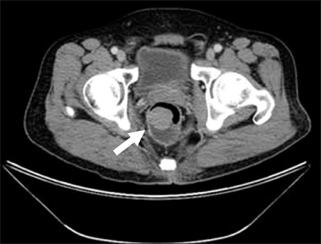
Fig. 1 Abdominopelvic computed tomography. A 6.5×3.3 cm intraluminal polypoid mass in the right lateral wall of the distal rectum was noted (arrow).

Fig. 2 Colonoscopic finding. A 3.3 cm sized subepithelial tumor in the rectum was noted.

Fig. 3 Endoscopic ultrasonography finding. A hypoechoic mass originating from the fourth endosonographic layer was noted.
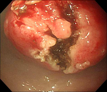
Fig. 4 Unroofing technique. Using a flex knife, direct biopsies were obtained after removing the overlying mucosa.

Fig. 5 (A) Low power view showed focal diffuse lymphoid infiltration (arrow; H&E, ×40). (B) Dense infiltration of centrocyte-like small lymphoid cells (H&E, ×400). (C) Lymphoid cells positive for CD20 by immunohistochemical staining (×400). (D) Lymphoid cells negative for cyclin D1 by immunohistochemical staining (×400).
Cited by 1 articles
-
Rectal Mucosa-associated Lymphoid Tissue Lymphoma Treated with Endoscopic Resection
Baek Hyun Yoon, Cheal Wung Huh
Korean J Gastroenterol. 2021;78(6):344-348. doi: 10.4166/kjg.2021.124.
Reference
-
1. Hwang JH, Rulyak SD, Kimmey MB. American Gastroenterological Association Institute. American Gastroenterological Association Institute technical review on the management of gastric subepithelial masses. Gastroenterology. 2006. 130:2217–2228.2. Lee CK, Chung IK, Lee SH, et al. Endoscopic partial resection with the unroofing technique for reliable tissue diagnosis of upper GI subepithelial tumors originating from the muscularis propria on EUS (with video). Gastrointest Endosc. 2010. 71:188–194.3. Yamamoto R, Kato S, Shimazaki K, et al. A case of primary rectal mucosa-associated lymphoid tissue lymphoma treated by endoscopic mucosal resection. Digestive Endoscopy. 2005. 17:172–174.4. Gavioli M, Bagni A, Santacroce G, Piccagli I, Natalini G. Endorectal sonographic appearances of rectal MALT lymphoma, its response to therapy, and local recurrence. J Clin Ultrasound. 2001. 29:401–405.5. Yatabe Y, Nakamura S, Nakamura T, et al. Multiple polypoid lesions of primary mucosa-associated lymphoid-tissue lymphoma of colon. Histopathology. 1998. 32:116–125.6. Tanaka S, Ohta T, Kaji E, Kosaka T, Murakami I. EMR of mucosa-associated lymphoid tissue lymphoma of the rectum. Gastrointest Endosc. 2003. 57:956–959.7. Isaacson P, Wright DH. Malignant lymphoma of mucosa-associated lymphoid tissue. A distinctive type of B-cell lymphoma. Cancer. 1983. 52:1410–1416.8. Harris NL, Jaffe ES, Stein H, et al. A revised European-American classification of lymphoid neoplasms: a proposal from the International Lymphoma Study Group. Blood. 1994. 84:1361–1392.9. Takenaka R, Tomoda J, Sakata T, et al. Mucosa-associated lymphoid tissue lymphoma of the rectum that regressed spontaneously. J Gastroenterol Hepatol. 2000. 15:331–335.10. Ahlawat S, Kanber Y, Charabaty-Pishvaian A, et al. Primary mucosa-associated lymphoid tissue (MALT) lymphoma occurring in the rectum: a case report and review of the literature. South Med J. 2006. 99:1378–1384.11. Mimura T, Kuramoto S, Hashimoto M, et al. Unroofing for lymphangioma of the large intestine: a new approach to endoscopic treatment. Gastrointest Endosc. 1997. 46:259–263.12. de la Serna-Higuera C, Pérez-Miranda M, Díez-Redondo P, et al. EUS-guided single-incision needle-knife biopsy: description and results of a new method for tissue sampling of subepithelial GI tumors (with video). Gastrointest Endosc. 2011. 74:672–676.
- Full Text Links
-
- Actions
-
Cited
- CITED
-
- Close
- Share
-
- Similar articles
-
- A Case of Primary Rectal Colon Mucosa associated Lymphoid Tissue Lymphoma
- Successful Endoscopic Resection of Residual Colonic Mucosa-Associated Lymphoid Tissue Lymphoma after Polypectomy
- Twin Rectal Tonsils Mimicking Carcinoid or Mucosa-Associated Lymphoid Tissue Lymphoma
- Rectal Mucosa-associated Lymphoid Tissue Lymphoma Treated with Endoscopic Resection
- Primary Biliary Mucosa-associated Lymphoid Tissue Lymphoma Mimicking Hilar Cholangiocarcinoma
