MALT (Mucosa-Associated Lymphoid Tissue) Lymphoma of the Colon
- Affiliations
-
- 1Department of Internal Medicine, Seoul Paik Hospital, Inje Univercity College of Medicine, Seoul, Korea. jw0412@korea.com
- KMID: 1792770
- DOI: http://doi.org/10.4166/kjg.2010.55.4.213
Abstract
- No abstract available.
MeSH Terms
Figure
-
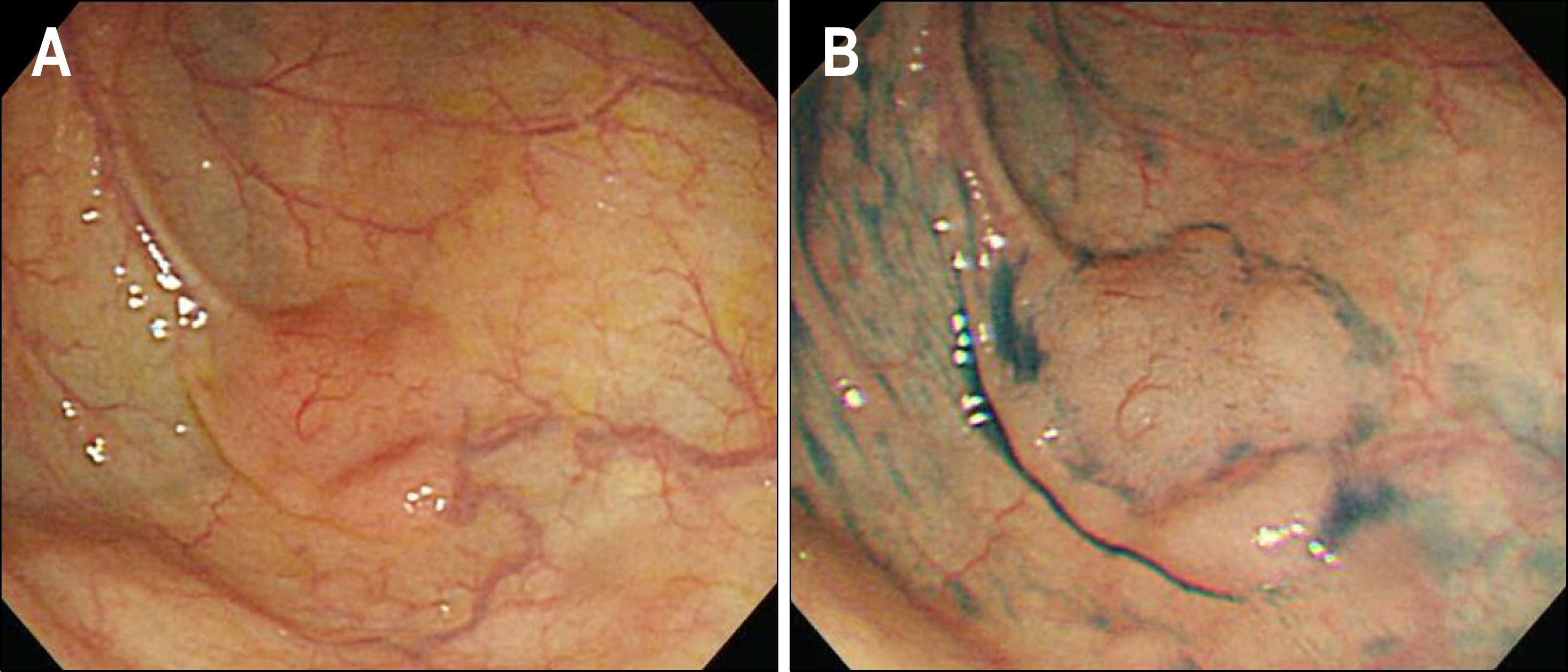
Fig. 1. Colonoscopic findings. (A) A single sessile polypoid lesion was noted on the cecum. (B) The lesion was observed more clearly after spraying indigocarmine.

Fig. 2. Histological findings. (A) Biopsy specimen showed diffuse infiltration of atypical lymphocytes in the mucosal layer (H&E, ×100). (B) Immunohistochemical stain for CD 20 was diffusely positive (×100), (C) and also stain for bcl-2 was positive (×100), (D) but stain for CD3 was negative (×100).

Fig. 3. Miniprobe EUS showed an 1.2 cm sized hypoechoic mass with focally disrupted submucoal and intact proper muscle layer of colon.
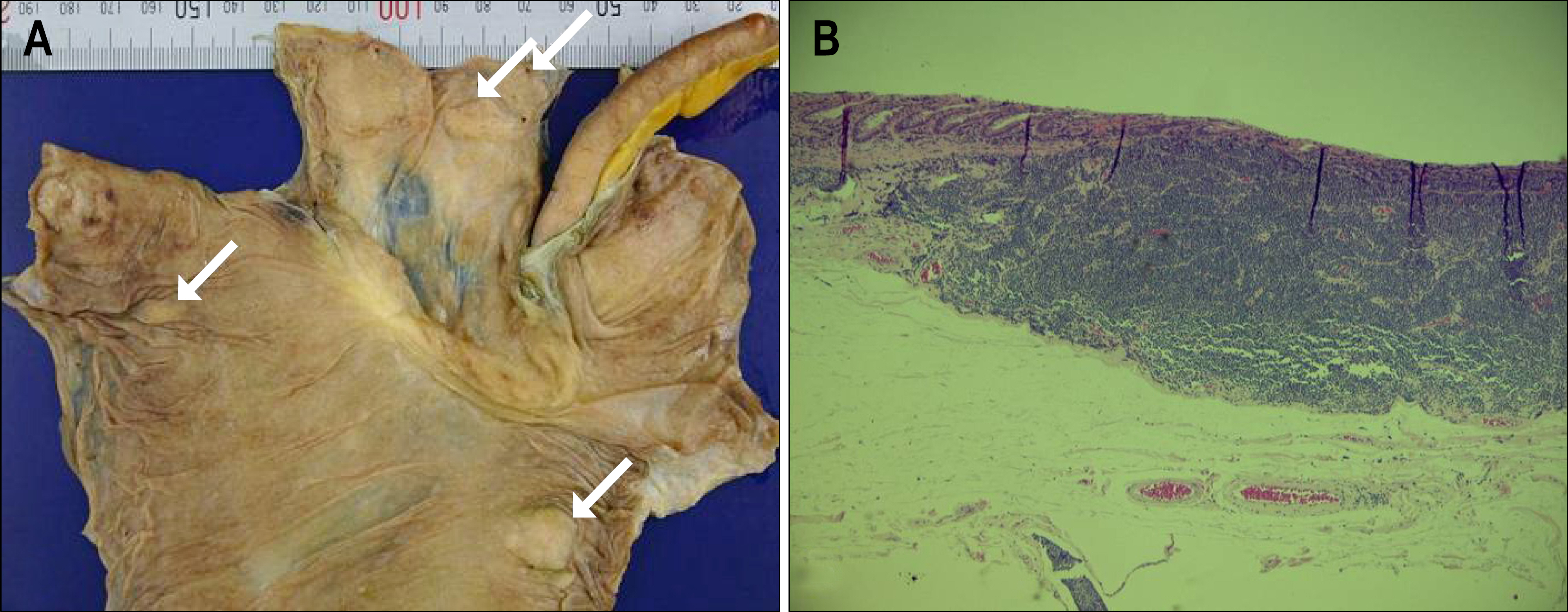
Fig. 4. Pathologic findings. (A) Gross specimen demonstrated multiple nodular lesions (arrows) on the cecum and terminal ileum. (B) The resection specimen showed diffuse infiltration of atypical lymphocytes in the mucosal and submucosal layer.
Cited by 1 articles
-
A Case of Mucosa-Associated Lymphoid Tissue Lymphoma of the Sigmoid Colon Presenting as a Semipedunculated Polyp
Myung Hwan Kim, Jin Tae Jung, Eui Jung Kim, Tae Won Kim, Seon Young Kim, Joong Goo Kwon, Eun Young Kim, Woo Jung Sung
Clin Endosc. 2014;47(2):192-196. doi: 10.5946/ce.2014.47.2.192.
Reference
-
1. Shepherd NA, Hall PA, Coates PJ, Levison DA. Primary malignant lymphoma of the colon and rectum. A histopathological and immunohistochemical analysis of 45 cases with clinicopathological correlations. Histopathology. 1988; 12:235–252.
Article2. Kahl BS. Update: gastric MALT lymphoma. Curr Opin Oncol. 2003; 15:347–352.
Article3. Cavalli F, Isaacson PG, Gascoyne RD, Zucca E. MALT lymphomas. Hematology Am Soc Hematol Educ Program. 2001. 241–258.
Article4. Hahn JS, Kim YS, Lee YC, Yang WI, Lee SY, Suh CO. Eleven-year experience of low grade lymphoma in Korea (based on REAL classification). Yonsei Med J. 2003; 44:757–770.
Article5. Wü ndisch T, Thiede C, Morgner A, et al. Longterm follow-up of gastric MALT lymphoma after Helicobacter pylori eradication. J Clin Oncol. 2005; 23:8018–8024.6. Shaye OS, Levine AM. Marginal zone lymphoma. J Natl Compr Canc Netw. 2006; 4:311–318.
Article7. Yatabe Y, Nakamura S, Nakamura T, et al. Multiple polypoid lesions of primary mucosa-associated lymphoid-tissue lymphoma of colon. Histopathology. 1998; 32:116–125.
Article8. Zucca E, Conconi A, Pedrinis E, et al. Nongastric marginal zone B-cell lymphoma of mucosa-associated lymphoid tissue. Blood. 2003; 101:2489–2495.
Article9. Rhee JC, Lee HY, Rhee PL, et al. Esophagus, Stomach & Intestine; endoscopic findings of gastric mucosa: associated lymphoid tissue (MALT) lymphoma. Korean J Gastrointest Endosc. 1997; 17:125–131.10. Schmid C, Vazquez JJ, Diss TC, Isaacson PG. Primary B-cell mucosa associated lymphoid tissue lymphoma presenting as a solitary colorectal polyp. Histopathology. 1994; 24:357–362.11. Chung CH, Kim HG, Park WS, et al. A case of mucosa-associated lymphoid tissue lymphoma of colon as multiple polypoid lesions. Korean J Gastrointest Endosc. 2001; 23:122–126.12. Lee YG, Lee S, Han SW, Lee JS. A case of multiple mucosa-associated lymphoid tissue (MALT) lymphoma of the colon identified as simple mucosal discoloration. J Korean Med Sci. 2005; 20:325–328.
Article
- Full Text Links
-
- Actions
-
Cited
- CITED
-
- Close
- Share
-
- Similar articles
-
- A Case of Mucosa-Associated Lymphoid Tissue Lymphoma of Colon as Multiple Large Polypoid Lesions
- A Case of Multiple Mucosa-Associated Lymphoid Tissue (MALT) Lymphoma of the Colon Identified as Simple Mucosal Discoloration
- A Case of Primary Rectal Colon Mucosa associated Lymphoid Tissue Lymphoma
- A case report of the Pulmonary Malignant Lymphomaof the mucosa-associated lymphoid tissue(MALT)
- A Case of Gastroduodenal Mucosa-associated Lymphoid Tissue Lymphoma Regression after Eradication of Helicobacter pylori
