Benign Schwannoma of the Liver: A Case Report
- Affiliations
-
- 1Department of Internal Medicine and Institute of Health Science, Gyeongsang National University School of Medicine, Jinju, Korea. kimthy@medimail.co.kr
- 2Department of Pathology, Gyeongsang National University School of Medicine, Jinju, Korea.
- KMID: 1785838
- DOI: http://doi.org/10.3346/jkms.2008.23.4.727
Abstract
- A primary benign schwannoma of the liver is extremely rare. Only nine cases have been reported in the medical literature worldwide and no case has been reported in Korea previously. A 36-yr-old woman was admitted to our hospital with vague epigastric pain. The ultrasound and computed tomography scan revealed a multiseptated cystic mass in the right lobe of the liver. The mass was resected; it was found to be a 5x4x2 cm mass filled with reddish yellow fluid. The histological examination confirmed the diagnosis of a benign schwannoma, proven by positive immunoreaction with the neurogenic marker S-100 protein and a negative response to CD34, CD117 and smooth muscle actin. This is the first report of a benign schwannoma of the liver parenchyma in a Korean patient.
Keyword
MeSH Terms
Figure
-
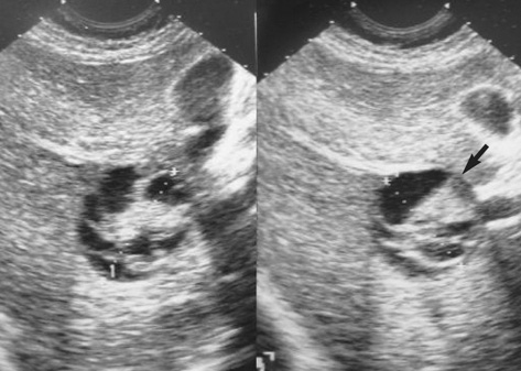
Fig. 1 Ultrasound findings of the mass. A 4.5-cm sized round hypoechoic mass with multiple central echogenic bands in the right lobe of the liver (segment V).
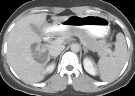
Fig. 2 Abdominal computed tomography shows a 5 cm smooth margined low-attenuating mass in the right lobe of liver (segment V), containing multiple central septations.
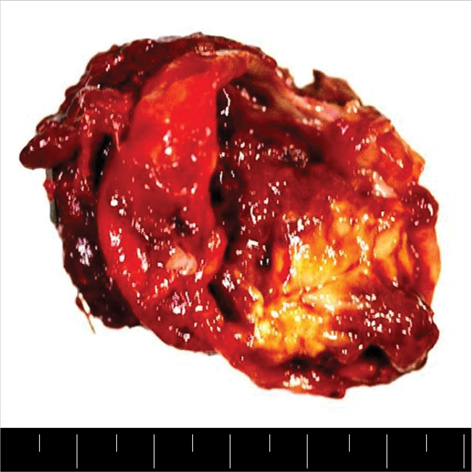
Fig. 3 A photograph showing the cut section of the tumor: The specimen was 5 × 4 × 2 cm. A multi-septated cyst that was previously opened, measuring 4 × 4 cm. The cyst was 1 cm from the resection margin. The cyst contained 10 soft tissues. The wall thickness measured 0.4 cm.
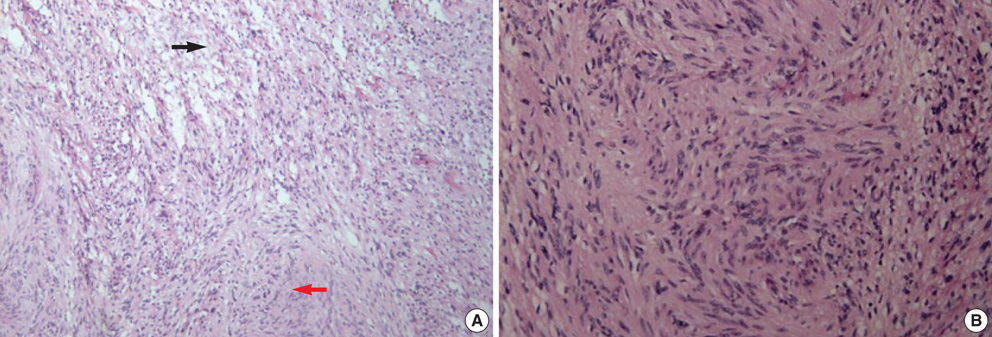
Fig. 4 (A) Spindle cell tumor with a mixture of two growth patterns, Antoni A and Antoni B, is seen. In the Antoni A pattern of growth, elongated cells were arranged in areas of moderate to high cellularity (black arrow). In the Antoni B pattern of growth, the tumor was less densely cellular, had a loose meshwork of cells, as well as microcysts and myxoids changes (red arrow) (H&E, ×100). (B) Spindle cell tumor with cells arranged in whorls in a storiform pattern is seen (H&E, ×200).
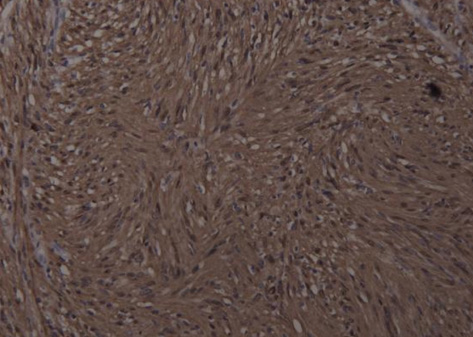
Fig. 5 A photograph of immunochemical staining with S-100 protein: The tumor is composed of bundles of compact spindle cells that are immunoreactive to the S-100 protein (×200).
Cited by 1 articles
-
Primary Hepatic Schwannoma
Youn I Choi, Yun Seob Kim, Ju Hyun Kim, Seong Hee Lee, Seong Gak Shin, Yun Soo Kim, Duck Joo Choi, Seung Joon Choi, Dong Hae Chung, Oh Sang Kwon
Korean J Gastroenterol. 2018;72(3):150-154. doi: 10.4166/kjg.2018.72.3.150.
Reference
-
1. Pereira Filho RA, Souza SA, Oliveira Filho JA. Primary neurilemmal tumour of the liver: case report. Arq Gastroenterol. 1978. 15:136–138.2. Bekker GM. Neurofibroma of the liver. Sov Med. 1982. 10:120–121.3. Hytiroglou P, Linton P, Klion F, Schwartz M, Miller C, Thung SN. Benign schwannoma of the liver. Arch Pathol Lab Med. 1993. 117:216–218.4. Heffron TG, Coventry S, Bedendo F, Baker A. Resection of primary schwannoma of the liver not associated with neurofibromatosis. Arch Surg. 1993. 128:1396–1398.
Article5. Yoshida M, Nakashima Y, Tanaka A, Mori K, Yamaoka Y. Benign schwannoma of the liver: a case report. Nippon Geka Hokan. 1994. 63:208–214.6. Wada Y, Jimi A, Nakashima O, Kojiro M, Kurohiji T, Sai K. Schwannoma of the liver: report of two surgical cases. Pathol Int. 1998. 48:611–617.
Article7. Flemming P, Frerker M, Klempnauer J, Pichlmayr R. Benign schwannoma of the liver with cystic changes misinterpreted as hydatid disease. Hepatogastroenterology. 1998. 45:1764–1766.8. Kapoor S, Tevatia MS, Dattagupta S, Chattopadhyay TK. Primary hepatic nerve sheath tumor. Liver Int. 2005. 25:458–459.
Article9. Park MK, Lee KT, Choi YS, Shin DH, Lee JY, Lee JK, Paik SW, Ko YH, Rhee JC. A case of benign schwannoma in the porta hepatis. Korean J Gastroenterol. 2006. 47:164–167.10. Ducatman BS, Scheithauer BW, Piepgras DG, Reiman HM, Ilstrup DM. Malignant peripheral nerve sheath tumors. A clinicopathologic study of 120 cases. Cancer. 1986. 57:2006–2021.
Article11. Brennan MF, Singer S, Maki RG, O'sullivan B. DeVita VT, Hellman S, editors. Soft tissue sarcoma. Cancer: Principles & practice of oncology. 2000. 7th edition. Philadelphia: Lippincott Williams & Wilkins;1595.12. Lantz PE, Listrom MB. Fenoglio-Preiser CM, Noffsinger AE, Stemmermann GN, editors. Gastrointestinal mesenchymal neoplasms. Gastrointestinal pathology. 2002. 2nd edition. Philadelphia: Lippincott-Raven;1185–1188.13. Williams PL, Warwick R, Dyson M, Bannister LH, editors. Gray's anatomy. 1989. 37th edition. New York: Churchill Livingstone;1165. 1390. 1395.14. Cohen LM, Schwartz AM, Rockoff SD. Benign schwannomas: pathologic basis for CT inhomogeneities. AJR Am J Roentgenol. 1986. 147:141–143.
Article15. Daimaru Y, Kido H, Hashimoto H, Enjoji M. Benign schwannoma of the gastrointestinal tract: a clinicopathologic and immunohistochemical study. Hum Pathol. 1988. 19:257–264.
Article16. Miettinen M, Lasota J. Gastrointestinal stromal tumors: review on morphology, molecular pathology, prognosis, and differential diagnosis. Arch Pathol Lab Med. 2006. 130:1466–1478.
Article17. Enzinger FM, Weiss SW. Enzinger FM, Weiss SW, editors. Benign tumors of peripheral nerves. Soft tissue tumors. 1995. 3rd edition. St. Louis: Mosby;829–842.
- Full Text Links
-
- Actions
-
Cited
- CITED
-
- Close
- Share
-
- Similar articles
-
- A case of cystic change of pelvic retroperitoneal Schwannoma misdiagnosed as an ovarian tumor
- A Case of Benign Retroperitoneal Schwannoma of the Obturator Fossa
- Benign Schwannoma Mimicking Metastatic Lesion on F-18 FDG PET/CT in Differentiated Thyroid Cancer
- Large Forefoot Schwannoma: A Case Report
- Benign Schwannoma of the Esophagus
