Intramedullary Clear Cell Ependymoma in the Thoracic Spinal Cord: A Case with Its Crush Smear and Ultrastructural Findings
- Affiliations
-
- 1Department of Pathology, Gachon University of Medicine and Science, Gil Medical Center, Incheon, Korea. syha@gilhospital.com
- 2Department of Neurosurgery, Gachon University of Medicine and Science, Gil Medical Center, Incheon, Korea.
- KMID: 1785805
- DOI: http://doi.org/10.3346/jkms.2007.22.S.S149
Abstract
- Clear cell ependymoma was included in the World Health Organization classification of the nervous system in 1993, and all the reported cases, except for two in the spinal cord, were located in the brain, mainly in the supratentorial compartment. Astrocytomas outnumber ependymomas in the spinal cord, and the two entities partly share cytologic findings such as long, bipolar glial processes and oval to round nuclei resembling those seen in pilocytic astrocytoma. Here, we report the first Korean case of intramedullary clear cell ependymoma of the spinal cord, which is the third case situated in the spinal cord in the literature. The crush smear revealed round-to-oval nuclei with occasional nuclear eosinophilic inclusion and rare nuclear grooves. Cytoplasm had fluffy eosinophilic glial processes, and acellular fibrillary zone. On hematoxylin-eosin stain, oval to round tumor cells had large central nuclei with indistinct nucleoli and a moderate amount of clear cytoplasm, i.e. perinuclear halo, mimicking oligodendroglioma. Perivascular pseudorosettes and ependymal clefts were rarely found. In retrospect, perinuclear halo was absent on crush smears. Ultrastructurally, they had extensive surface microvilli and edematous cytoplasm filled with abundant glial filaments and microlumens with or without microvilli. Intercellular long cell junctions of the zipper-like zonula adherens type were found.
MeSH Terms
Figure
-
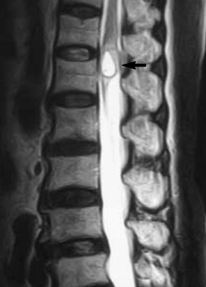
Fig. 1 MR images of the thoracic spinal cord. A T1-weighted image demonstrates that the tumor is isodense (arrow).
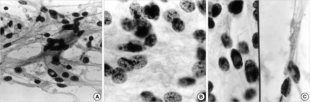
Fig. 2 Touch preparation on H&E stain. Smear preparation with long glial processes and oval to round tumor nuclei. (A) Tumor cells show fibrillary cytoplasm and euchromatic round to vesicular nuclei. (B) Acellular fibrillary zones. (C) Intranuclear pseudoinclusions (left) and nuclear grooves (right).
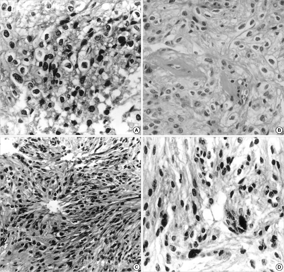
Fig. 3 H&E stain. (A, B) Clear cells with round nuclei with perinuclear halo and eosinophilic cytoplasm. (C) Ependymal rosettes. (D) Nuclear pleomorphism.

Fig. 4 (A) The tumor cells show abundant cytoplasmic long processes containing intermediate filaments, microtubules, free ribosomes, rough endoplasmic reticulums (×5,400). (B) Cell junctions of zonula adherens type and abundant microvilli with rare cilia are found (×27,000).
Reference
-
1. Kawano N, Yada K, Aihara M, Yagishita S. Oligodendroglioma-like cells (clear cells) in ependymoma. Acta Neuropathol (Berl). 1983. 62:141–144.
Article2. Fouladi M, Helton K, Dalton J, Gilger E, Gajjar A, Merchant T, Kun L, Newsham I, Burger P, Fuller C. Clear cell ependymoma: a clinicopathologic and radiographic analysis of 10 patients. Cancer. 2003. 98:2232–2244.
Article3. Kim YJ, Tsunoda S, Yokoyama K, Miyamoto K, Tamai M, Yamauchi M. Clear cell ependymoma with a lipidized component that developed in the thoracic spinal cord. Neurol Res. 2003. 25:324–328.
Article4. Akutsu H, Shibata Y, Okazaki M, Hyodo A, Matsumura A. Intramedullary clear cell ependymoma in the cervical spinal cord: case report. Neurosurgery. 2000. 47:1434–1437.
Article5. Chamberlain MC. Ependymomas. Curr Neurol Neurosci Rep. 2003. 3:193–199.
Article6. Zulch KJ. Brain tumours. Their biology and pathology. 1957. 1st ed. New York, USA: Springer-Verlag.7. Min KW, Scheithauer BW. Clear cell ependymoma: a mimic of oligodendroglioma: clinicopathologic and ultrastructural considerations. Am J Surg Pathol. 1997. 21:820–826.
Article8. Kawano N, Ohba Y, Nagashima K. Eosinophilic inclusions in ependymoma represent microlumina: a light and electron microscopic study. Acta Neuropathol. 2000. 99:214–218.
Article9. Kumar PV. Nuclear grooves in ependymoma. Cytologic study of 21 cases. Acta Cytol. 1997. 41:1726–1731.10. Amatya VJ, Takeshima Y, Kaneko M, Nakano T, Yamaguchi S, Sugiyama K, Kurisu K, Nakazato Y, Inai K. Case of clear cell ependymoma of medulla oblongata: clinicopathological and immunohistochemical study with literature review. Pathol Int. 2003. 53:297–302.
Article
- Full Text Links
-
- Actions
-
Cited
- CITED
-
- Close
- Share
-
- Similar articles
-
- Crush Cytologic Findings of Myxopapillary Ependymoma in Spinal Cord: A Case Report
- Thoracic Intramedullay Clear Cell Meningioma
- Clinical Outcome and Follow-up of Neonatal Hydronephrosis Diagnosed Antenatally
- A Case of Intramedullary Ependymoma in the upper Thoracic Spinal Cord
- A Case of Spinal Cord Ependymoma
