J Korean Med Sci.
2007 Sep;22(Suppl):S61-S65. 10.3346/jkms.2007.22.S.S61.
The Characteristics of Incidental Pituitary Microadenomas in 120 Korean Forensic Autopsy Cases
- Affiliations
-
- 1Department of Pathology, Ajou University School of Medicine, Suwon, Korea. kyibeom@ajou.ac.kr
- 2Division of Forensic Medicine, National Institute of Scientific Investigation, Seoul, Korea.
- KMID: 1785790
- DOI: http://doi.org/10.3346/jkms.2007.22.S.S61
Abstract
- To investigate the characteristics of incidental pituitary microadenomas, we examined 120 pituitary glands from Korean forensic autopsy cases, from which eight tumors were identified (incidence 6.7%). The average age of the affected subjects was 50 yr (range: 33-96 yr) with a female predominance. The maximum diameters of the tumors ranged from 0.4 to 5.4 mm (mean: 2.8 mm). Immunohistochemical analysis of pituitary hormones revealed three growth hormone-secreting adenomas, one prolactin-producing adenoma, one gonadotropin-producing adenoma, one plurihormonal adenoma, and two null cell adenomas. MIB-1 staining for Ki-67 antigen showed no positive expression. The microvessel density (MVD) of the pituitary microadenomas ranged from 2.3 to 11.6% (mean: 5.3%) and was significantly lower than that of nonneoplastic pituitary glands (11.9-20.1%, mean: 14.8%). Our study provides reference data on incidental pituitary microadenomas in the Korean population.
MeSH Terms
Figure
-

Fig. 1 (A) Pituitary microadenoma (H&E stain, ×100). (B) Loss of the normal acinar and stromal patterns and compression of the adjacent pituitary parenchyma (reticulin stain, ×100). (C) Positive immunoreactivity with prolactin (×100).

Fig. 2 Microvessel density measurement using an image analyzer with CD 34 immunostain (×200).
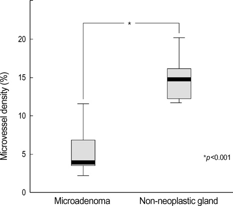
Fig. 3 Microvessel density in microadenomas and non-neoplastic pituitary gland showing a significant difference.
Reference
-
1. Krikorian A, Aron D. Evaluation and management of pituitary incidentalomas--revisiting an acquaintance. Nat Clin Pract Endocrinol Metab. 2006. 2:138–145.
Article2. Aron DC, Howlett TA. Pituitary incidentalomas. Endocrinol Metab Clin North Am. 2000. 29:205–221.
Article3. Sanno N, Oyama K, Tahara S, Teramoto A, Kato Y. A survey of pituitary incidentaloma in Japan. Eur J Endocrinol. 2003. 149:123–127.
Article4. Molitch ME, Russell EJ. The pituitary "incidentaloma". Ann Intern Med. 1990. 112:925–931.
Article5. Arita K, Tominaga A, Sugiyama K, Eguchi K, Iida K, Sumida M, Migita K, Kurisu K. Natural course of incidentally found nonfunctioning pituitary adenoma, with special reference to pituitary apoplexy during follow-up examination. J Neurosurg. 2006. 104:884–891.
Article6. Camaris C, Balleine R, Little D. Microadenomas of the human pituitary. Pathology. 1995. 27:8–11.
Article7. Parent AD, Brown B, Smith EE. Incidental pituitary adenomas: a retrospective study. Surgery. 1982. 92:880–883.8. Burrow GN, Wortzman G, Rewcastle NB, Holgate RC, Kovacs K. Microadenomas of the pituitary and abnormal sellar tomograms in an unselected autopsy series. N Engl J Med. 1981. 304:156–158.
Article9. Fainstein Day P, Guitelman M, Artese R, Fiszledjer L, Chervin A, Vitale NM, Stalldecker G, De Miguel V, Cornalo D, Alfieri A, Susana M, Gil M. Retrospective multicentric study of pituitary incidentalomas. Pituitary. 2004. 7:145–148.
Article10. Gorczyca W, Hardy J. Microadenomas of the human pituitary and their vascularization. Neurosurgery. 1988. 22:1–6.
Article11. Parent AD, Bebin J, Smith RR. Incidental pituitary adenomas. J Neurosurg. 1981. 54:228–231.
Article12. Tomita T, Gates E. Pituitary adenomas and granular cell tumors. Incidence, cell type, and location of tumor in 100 pituitary glands at autopsy. Am J Clin Pathol. 1999. 111:817–825.
Article13. Kastelan D, Korsic M. High prevalence rate of pituitary incidentaloma: Is it associated with the age-related decline of the sex hormones levels? Med Hypotheses. 2007. 69:307–309.
Article14. Char G, Persaud V. Asymptomatic microadenomas of the pituitary gland in an unselected autopsy series. West Indian Med J. 1986. 35:275–279.15. Feldkamp J, Santen R, Harms E, Aulich A, Modder U, Scherbaum WA. Incidentally discovered pituitary lesions: high frequency of macroadenomas and hormone-secreting adenomas-results of a prospective study. Clin Endocrinol (Oxf). 1999. 51:109–113.16. DeStephano DB, Lloyd RV, Pike AM, Wilson BS. Pituitary adenomas. An immunohistochemical study of hormone production and chromogranin localization. Am J Pathol. 1984. 116:464–472.17. Paek KI, Kim SH, Song SH, Choi SW, Koh HS, Youm JY, Kim Y. Clinical significance of Ki-67 labeling index in pituitary macroadenoma. J Korean Med Sci. 2005. 20:489–494.
Article18. Vidal S, Kovacs K, Horvath E, Scheithauer BW, Kuroki T, Lloyd RV. Microvessel density in pituitary adenomas and carcinomas. Virchows Arch. 2001. 438:595–602.
Article19. Yonezawa K, Tamaki N, Kokunai T. Clinical features and growth fractions of pituitary adenomas. Surg Neurol. 1997. 48:494–500.
Article20. Losa M, Barzaghi RL, Mortini P, Franzin A, Mangili F, Terreni MR, Giovanelli M. Determination of the proliferation and apoptotic index in adrenocorticotropin-secreting pituitary tumors: comparison between micro- and macroadenomas. Am J Pathol. 2000. 156:245–251.21. Niveiro M, Aranda FI, Peiro G, Alenda C, Pico A. Immunohistochemical analysis of tumor angiogenic factors in human pituitary adenomas. Hum Pathol. 2005. 36:1090–1095.
Article22. Turner HE, Nagy Z, Gatter KC, Esiri MM, Harris AL, Wass JA. Angiogenesis in pituitary adenomas and the normal pituitary gland. J Clin Endocrinol Metab. 2000. 85:1159–1162.
Article23. Kristof RA, Aliashkevich AF, Hans V, Haun D, Meyer B, Thees C, Schramm J. The regional oxygen saturation of pituitary adenomas is lower than that of the pituitary gland: microspectrophotometric study with potential clinical implications. Neurosurgery. 2003. 53:880–885.
Article24. Vidal S, Horvath E, Kovacs K, Lloyd RV, Scheithauer BW. Microvascular structural entropy: a novel approach to assess angiogenesis in pituitary tumors. Endocr Pathol. 2003. 14:239–247.
Article
- Full Text Links
-
- Actions
-
Cited
- CITED
-
- Close
- Share
-
- Similar articles
-
- Profile of Forensic Autopsy Practices in Small Towns and Cities in Rural Areas
- Forensic Pathology of the Lung in Fetus and Infant
- Examination of Abdominal Organ in Forensic Autopsy
- Incidental Superior Hypophygeal Artery Aneurysm Embedded within Pituitary Adenoma
- Sudden Death associated with Thyrotoxicosis: Report of Three Autopsy Cases
