Usefulness of Pain Distribution Pattern Assessment in Decision-Making for the Patients with Lumbar Zygapophyseal and Sacroiliac Joint Arthropathy
- Affiliations
-
- 1Department of Neurosurgery, Presbyterian Medical Center, Jeonju, Korea. hyoungihl@hotmail.com
- 2Department of Neurosurgery, College of Medicine, Yonsei University, Seoul, Korea.
- KMID: 1785768
- DOI: http://doi.org/10.3346/jkms.2007.22.6.1048
Abstract
- There are currently no initial guides for the diagnosis of somatic referred pain of lumbar zygapophyseal joint (LZJ) or sacroiliac joint (SIJ). We developed a classification system of LZJ and SIJ pain, the "pain distribution pattern template (PDPT)" depending on the pain distribution patterns from a pool of 200 patients whose spinal pain source was confirmed. We prospectively applied the PDPT to determine its contribution to clinical decision-making for 419 patients whose pain was presumed to arise from the LZJs (259 patients) or SIJs (160 patients). Forty-nine percent (128/259) of LZJ and 46% (74/160) of SIJ arthopathies diagnosed by PDPT were confirmed by nerve blocks. Diagnostic reliabilities were significantly higher in Type A and C patterns in LZJ and Type C in SIJ arthropathies, 64%, 80%, and 68.4%, respectively. For both LZJ and SIJ arthropathies, favorable outcome after radiofrequency (RF) neurotomies was similar to the rate of positive responses to diagnostic blocks in Type A to Type D, whereas the outcome was unpredictable in those with undetermined type (Type E). Considering the paucity of currently available diagnostic methods for LZJ and SIJ arthropathies, PDPT is useful in clinical decision- making as well as in predicting the treatment outcome.
MeSH Terms
Figure
-
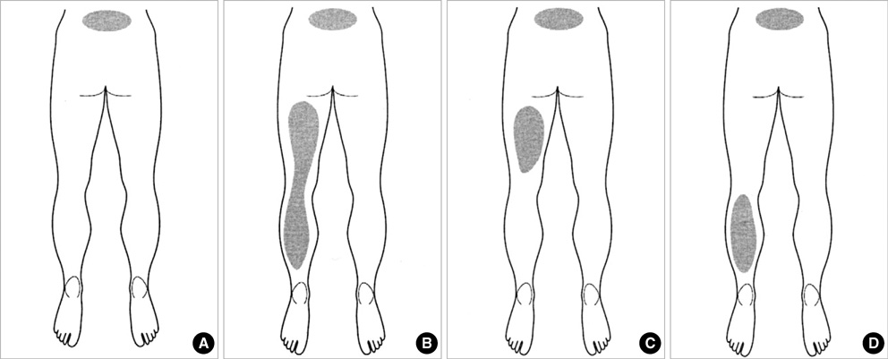
Fig. 1 Types of pain distribution patterns in lumbar zygapophyseal joint arthropathy. Type A shows the pain distribution in the paravertebral area localized or band-like extension; type B combines type A and leg pain in the posterior thigh and calf; type C combines type A and leg pain in the posterior thigh only; type D demonstrates the combination of type A and leg pain confined to calf; type E, not depicted here, is the undetermined type, which does not belong to the above types.
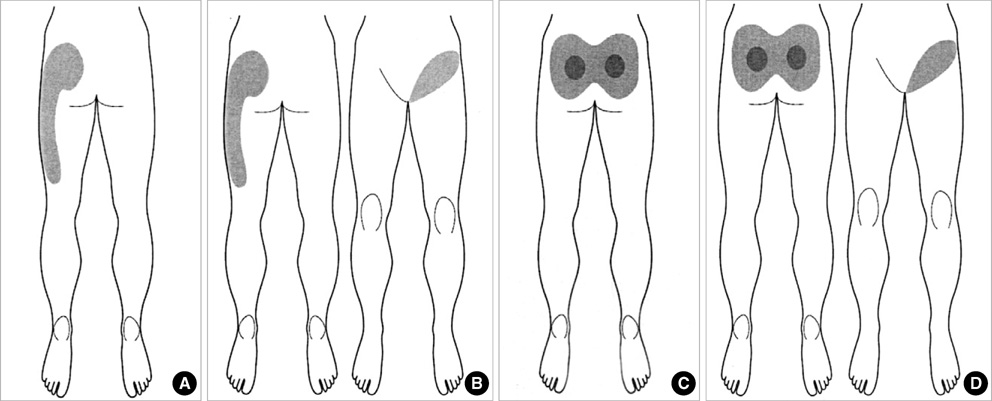
Fig. 2 Types of pain distribution patterns in sacroiliac joint arthropathy. Type A has pain distribution in the lower back, gluteal region, and lateral aspect of the thigh; Type B is similar to type A but with the addition of groin pain; Type C has pain in the lower back and posterior aspects of the gluteal region; Type D combines type C and groin pain; Type E, not depicted here, indicates the undetermined pattern.

Fig. 3 Treatment effect depending upon the pain distribution patterns in lumbar zygapophyseal joint (A) and sacroiliac joint arthropathy (B). Favorable outcomes in nerve blocks were correlated with that of RF procedures. However, patients in type E of lumbar zygapophyseal and sacroiliac joint arthropathies showed better results from the RF procedure, despite lower rates of favorable outcomes in nerve block.
Cited by 2 articles
-
Non-operative spinal interventions for cervical and lumbar spinal pain
Jaemin Lee
J Korean Med Assoc. 2014;57(4):297-299. doi: 10.5124/jkma.2014.57.4.297.Nonoperative interventions for spinal pain
Dong Ah Shin, Hyoung Ihl Kim
J Korean Med Assoc. 2014;57(4):308-317. doi: 10.5124/jkma.2014.57.4.308.
Reference
-
1. Nachemson AL, Wadell G, Norlund AI. Nachemson AL, Jonsson E, editors. Epidemiology of neck and low back pain. Neck and back pain: the scientific evidence causes, diagnosis, and treatment. 2000. Philadelphia: Lippincott, Williams and Wilkins;165–188.2. Andersson GB. Frymoyer JW, editor. The epidemiology of spinal disorders. The adult spine: principles and practice. 1997. Philadelphia: Lippincott-Raven;93–141.3. Deyo RA, Tsui-Wu YJ. Descriptive epidemiology of low back pain and its related medical care in the United States. Spine. 1987. 12:264–268.4. Bogduk N. Bogduk N, editor. Low back pain. Clinical anatomy of the lumbar spine and sacrum. 2002. London: Churchill Livingstone;187–213.5. Boos N, Rieder R, Schade V, Psych D, Spratt KF, Semmer N, Psych D, Aebi M. The diagnostic accuracy of magnetic resonance imaging, work perception, and psychosocial factors in identifying symptomatic disc herniations. Spine. 1995. 20:2613–2625.
Article6. Modic MT, Ross JS, Obuchowski NA, Browning KH, Cianflocco AJ, Mazanec DJ. Contrast-enhanced MR imaging in acute lumbar radiculopathy: a pilot study of the natural history. Radiology. 1995. 195:429–435.
Article7. Kim HI, Shin DG, Shin DA, Lee JO. Pain evaluation for decision making for management of spinal pain. J Korean Soc Ster Func Neurosurg. 2005. 1:44–50.8. Dreyfuss P, Michaelsen M, Pauza K, Mclarty J, Bogduk N. The value of medical history and physical examination in diagnosing sacroiliac joint pain. Spine. 1996. 21:2594–2602.
Article9. Slipman CW, Sterenfeld EB, Chou LH, Herzog R, Vresilovic E. The predictive value of provocative sacroiliac joint stree maneuvers in the diagnosis of sacroiliac joint syndrome. Arch Phys Med Rehabil. 1998. 79:288–292.10. Benneker LM, Heini PF, Anderson SE, Alini M, Ito K. Correlation of radiographic and MRI parameters to morphological and biochemical assessment of intervertebral disc degeneration. Eur Spine J. 2005. 14:27–35.
Article11. Finch P. Technology Insight: imaging of low back pain. Nat Clin Pract Rheumatol. 2006. 2:554–561.
Article12. Schwarzer AC, Wang SC, O'Driscoll D, Harrington T, Bogduk N, Laurent R. The ability of computed tomography to identify a painful zygapophysial joint in patient with chronic low back pain. Spine. 1995. 20:907–912.13. Yin W, Willard F, Carreiro J, Dreyfuss P. Sensory stimulation-guided sacroiliac joint radiofrequency neurotomy: technique based on neuroanatomy of the dorsal sacral plexus. Spine. 2003. 28:2419–2425.
Article14. Kim HJ, Shin DG, Kim HI, Shin DA. Selective neurotomy of sacral lateral branches for pain of sacroiliac joint dysfunction. J Korean Neurosurg Soc. 2005. 38:338–343.15. Carnes D, Ashby D, Underwood M. A systematic review of pain drawing literature: should pain drawings be used for psychologic screening? Clin J Pain. 2006. 22:449–457.16. Pande KC, Tripathi S, Kanoi R. Limited clinical utility of pain drawing in assessing patients with low back pain. J Spinal Disord Tech. 2005. 18:160–162.
Article17. Grunnesjo M, Bogefeldt J, Blomberg S, Delaney H, Svardsudd K. The course of pain drawings during a 10-week treatment period in patients with acute and sub-acute low back pain. BMC Musculoskelet Disord. 2006. 7:65.
Article18. Lyle MA, Manes S, McGuinness M, Ziaei S, Iversen MD. Relationship of physical examination findings and self-reported symptom severity and physical function in patients with degenerative lumbar conditions. Phys Ther. 2005. 85:120–133.
Article19. Slipman CW, Plastaras CT, Palmitier RA, Huston CW, Sterenfeld EB. Symptom provocation of fluoroscopically guided cervical nerve root stimulation: are dynatomal maps identical to dermatomal maps? Spine. 1998. 23:2235–2242.20. O'Neill CW, Kurgansky ME, Derby R, Ryan DP. Disc stimulation and patterns of referred pain. Spine. 2002. 27:2776–2781.21. Reigo T, Tropp H, Timpka T. Pain drawing evaluation--the problem with the clinically biased surgeon. Intra- and interobserver agreement in 50 cases related to clinical bias. Acta Orthop Scand. 1998. 69:408–411.
Article22. Gray DP, Bajwa ZH, Warfield CA. Waldman SD. Facet block and neurolysis. Interventional pain Management. 2001. 2nd. Philadephia, London: W.B. Saunders Company;446–483.23. Schwarzer AC, Aprill CN, Derby R, Fortin J, Kine G, Bogduk N. The relative contributions of the disc and zygapophyseal joint in chronic low back pain. Spine. 1994. 19:801–806.
Article24. Schwarzer AC, Aprill CN, Bogduk N. The sacroiliac joint in chronic low back pain. Spine. 1995. 20:31–37.
Article25. Bogduk N, Holmes S. Controlled zygapophysial joint blocks: the travesty of cost-effectiveness. Pain Med. 2000. 1:24–34.
Article26. Cihangiroglu M, Yildirim H, Bozgeyik Z, Senol U, Ozdemir H, Topsakal C, Yilmaz S. Observer variability based on the strength of MR scanners in the assessment of lumbar degenerative disc disease. Eur J Radiol. 2004. 51:202–208.
Article27. Dreyfuss P, Tibiletti C, Dreyer SJ. Thoracic zygapophyseal joint pain patterns.A study in normal volunteers. Spine. 1994. 19:807–811.28. McCall IW, Park WM, O'Brien JP. Induced pain referral from posterior lumbar elements in normal subjects. Spine. 1979. 441–446.
Article29. Suseki K, Takahashi Y, Takahashi K, Chiba T, Tanaka K, Morinaga T, Nakamura S, Moriya H. Innervation of the lumbar facet joints: origins and functions. Spine. 1997. 22:477–485.30. Bogduk N, Engel R. The menisci of the lumbar zygapophysial joints: A review of their anatomy and clinical significance. Spine. 1984. 9:454–460.
