Glial Choristoma in the Middle Ear and Mastoid Bone: A Case Report
- Affiliations
-
- 1Department of Pathology, Otolaryngology, College of Medicine, Dongguk University, Gyeongju, Korea. leego@mail.dongguk.ac.kr
- 2Department of Otolaryngology, College of Medicine, Dongguk University, Gyeongju, Korea.
- 3Department of Neurology, College of Medicine, Dongguk University, Gyeongju, Korea.
- KMID: 1785714
- DOI: http://doi.org/10.3346/jkms.2004.19.1.155
Abstract
- Heterotopic brain tissue usually involves extracranial midline structures of the head and neck such as nose, nasopharynx, and oral cavity. Its occurrence in the non-midline structures, including middle ear, is rare. We described a 50-yr-old-man with heterotopic glial tissue in the middle ear and mastoid bone. The patient presented with progressive hearing loss for 8 yr. There was no history of congenital anomalies, trauma, or ear surgery. Computed tomography revealed a mass-like lesion with soft tissue density occupying the middle ear cavity and mastoid antrum. At the operation, a graywhite fibrotic mass was detected in the epitympanic area. Mesotympanum and ossicles were intact. The patient underwent left simple mastoidectomy with type I tympanoplasty. During operation, definite cranial bone defect or cerebrospinal fluid leakage was not found. Histologically, the lesion was composed of exclusively mature, disorganized glial tissue with fibrovascular elements in a rather loose fibrillary background. Glial tissue showed diffuse positive reaction for glial fibrillar acidic protein and S100 protein on immunohistochemical study.
Keyword
MeSH Terms
Figure
-

Fig. 1 Preoperative pure tone audiometry shows conductive hearing loss on the left ear (10/70 dB). Right ear is normal (7/10 dB).

Fig. 2 Coronal (A, B) and axial (C, D) computed tomography demonstrate a mass-like lesion with soft tissue density occupying the middle ear and mastoid cavity of the left ear (B, D).
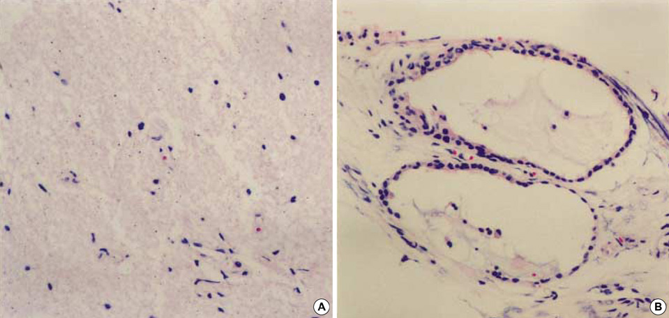
Fig. 3 Histologic features of the lesion. (A) The lesion is composed of exclusively mature disorganized glial tissue and fibrovascular elements in a rather loose fibrillary background. (B) Distended glandular structures are noted at the periphery of the glial tissue (H&E, ×100).
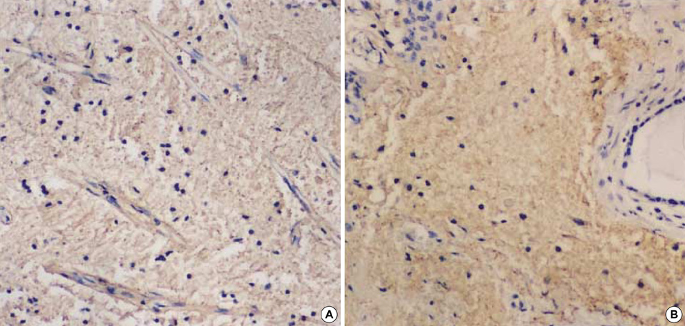
Fig. 4 Immunohistochemical features. The lesion shows diffuse positive reaction for GFAP (A) and S100 protein (B) (×100).
Cited by 1 articles
-
Glial Choristoma Accompanied with Severe Bony Defect in Orbit in a Newborn
Suk Hwan Han, Tae Hee Kim, Na Mi Lee, Jae Kyun Kim, She Young Lee
Korean J Perinatol. 2014;25(4):307-310. doi: 10.14734/kjp.2014.25.4.307.
Reference
-
1. Klein MV, Schwaighofer BW, Sobel DF, Fantozzi RD, Hesselink JR. Heterotopic brain in the middle ear: CT findings. J Comput Assist Tomogr. 1989. 13:1058–1060.2. Yeoh GP, Bale PM, de Silva M. Nasal cerebral heterotopia: the so-called nasal glioma or sequestered encephalocele and its variants. Pediatr Pathol. 1989. 9:531–549.
Article3. Choi HJ, Lee YS, Kim YS, Kim KY, Kang CS, Shim SI. Heterotopic brain tissue in the soft palate. Korean J Pathol. 1998. 32:1039–1041.4. Abdelsayed RA, Wetherington RW, Bent JP 3rd, Sharpe DE. Glial choristoma of the tongue: a case report and review of the literature. Oral Surg Oral Med Oral Pathol Oral Radiol Endod. 1999. 87:215–222.5. Ha SL, Shin JE, Yoon TH. Salivary gland choristoma of the middle ear: a case report. Am J Otolaryngol. 2000. 21:127–130.
Article6. Buckmiller LM, Brodie HA, Doyle KJ, Nemzek W. Choristoma of the middle ear: a component of a new syndrome? Otol Neurotol. 2001. 22:363–368.
Article7. Gyure KA, Thompson LD, Morrison AL. A clinicopathological study of 15 patients with neuroglial heterotopias and encephaloceles of the middle ear and mastoid region. Laryngoscope. 2000. 110:1731–1735.
Article8. McGregor DH, Cherian R, Kepes JJ, Kepes M. Case reports: heterotopic brain tissue of middle ear associated with cholesteatoma. Am J Med Sci. 1994. 308:180–183.9. Gulya AJ, Glasscock ME 3rd, Pensak ML. Neural choristoma of the middle ear. Otolaryngol Head Neck Surg. 1987. 97:52–56.
Article10. Slater DN, Timperley WR, Smith CM. Heterotopic middle ear gliomatosis. Histopathology. 1988. 12:230–231.
Article11. Wolbach SB. Congenital rhabdomyoma of the heart. Report of a case associated with multiple nests of neuroglia tissue in the meninges of the spinal cord. J Med Res. 1907. 16:495–520.12. Harris CP, Townsend JJ, Klatt EC. Accessory brains (extracerebral heterotopias): unusual prenatal intracranial mass lesions. J Child Neurol. 1994. 9:386–389.
Article13. Gyure KA, Morrison AL, Jones RV. Intracranial extracerebral neuroglial heterotopia: a case report and review of the literature. Ann Diagn Pathol. 1999. 3:182–186.
Article14. Whittet HB, Barker S, Anslow P, Leighton S. Heterotopic brain tissue: a rare cause of adult recurrent meningitis. J Laryngol Otol. 1990. 104:328–330.
Article15. Genut AA, Miranda FG, Garcia JH. Organized cerebral heterotopia in the ethmoid sinus. A case report. J Neurol Sci. 1976. 28:339–344.
- Full Text Links
-
- Actions
-
Cited
- CITED
-
- Close
- Share
-
- Similar articles
-
- Glial Choristoma of the Middle Ear: A Case Report
- Glial Choristoma Accompanied with Severe Bony Defect in Orbit in a Newborn
- Neuroglial Choristoma of the Middle Ear with Massive Tympanosclerosis: A Case Report and Literature Review
- Bilateral Bifurcation of the Mastoid Segment of the Facial Nerve
- Congenital Cholesteatoma Localized to the Tip of the Mastoid Bone: A Case Report and Possible Etiology
