J Korean Med Sci.
2004 Feb;19(1):149-151. 10.3346/jkms.2004.19.1.149.
Invasive Cystic Hypersecretory Carcinoma of the Breast: A Case Report
- Affiliations
-
- 1Department of Pathology, Seonam Univerisy, College of Medicine, Namwon, Korea. jshinlee@hanmail.net
- 2Department of Pathology, St. Carollo Hospital, Suncheon, Korea.
- KMID: 1785712
- DOI: http://doi.org/10.3346/jkms.2004.19.1.149
Abstract
- Cystic hypersecretory lesions of the breast are rare. These breast lesions include cystic hypersecretory hyperplasia (CHH), atypical CHH, and cystic hypersecretory carcinoma (CHC). The characteristic features are dilated ducts and cysts filled with thyroid colloid-like eosinophilic secretion. Only seven cases of invasive CHC have been reported in the literature. Here, we report an additional case of invasive CHC. The histologic features of the tumor showed both micropapillary intraductal carcinoma and focal high-grade invasive carcinoma in a background of CHH. This case suggests that cystic hypersecretory breast lesions encompass a spectrum of pathologic lesions including CHH, atypical CHH, CHC, and invasive CHC.
Keyword
MeSH Terms
Figure
-
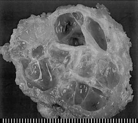
Fig. 1 The cut surface of the mass shows numerous cysts, measuring up to 1.2 cm in diameter, with a gelatinous secretion.
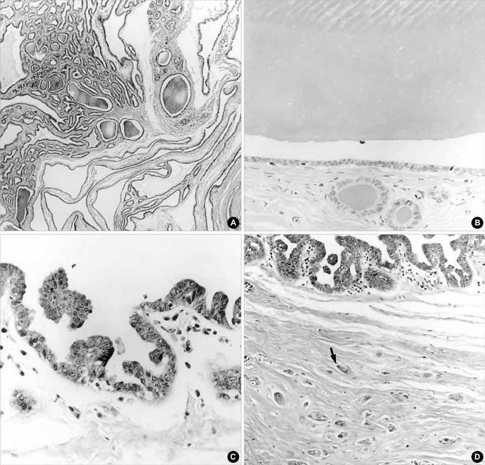
Fig. 2 Microscopic findings. The lesion is composed of multiple cysts and ducts containing eosinophilic secretion (A, H&E, ×20). Most of the cysts are lined by flat epithelium. The secretion retracts from the surrounding epithelium (B, H&E, ×200). The epithelium of some cysts grows as micropapillary intraductal carcinoma (C, H&E, ×200). An invasive component (arrow) is found adjacent to the intraductal carcinoma component (D, H&E, ×100).
Reference
-
1. Rosen PP, Scott M. Cystic hypersecretory duct carcinoma of the breast. Am J Surg Pathol. 1984. 8:31–41.
Article2. Guerry P, Erlandson RA, Rosen PP. Cystic hypersecretory hyperplasia and cystic hypersecretory duct carcinoma of the breast: pathology, therapy, and follow-up of 39 patients. Cancer. 1988. 61:1611–1620.3. Kim MK, Kwon GY, Gong GY. Fine needle aspiration cytology of cystic hypersecretory carcinoma of the breast: a case report. Acta Cytol. 1997. 41:892–896.4. Herrmann ME, McClatchey KD, Siziopikou KP. Invasive cystic hypersecretory ductal carcinoma of breast: a case report and review of the literature. Arch Pathol Lab Med. 1999. 123:1108–1110.5. Lee WY, Cheng L, Chang TW. Diagnosing invasive cystic hypersecretory duct carcinoma of the breast with fine needle aspiration cytology: a case report. Acta Cytol. 1999. 43:273–276.6. Shah AK, Banerjee SN, Sehonanda AS, Girishkumar H, Gerst PH. Cystic hypersecretory duct carcinoma of the breast. Breast J. 2000. 6:269–272.
Article7. Park JM, Seo MR. Cystic hypersecretory duct carcinoma of the breast: report of two cases. Clin Radiol. 2002. 57:312–315.
Article8. Lamovec J, Bracko M. Secretory carcinoma of the breast: light microscopical, immunohistochemical and flow cytometric study. Mod Pathol. 1994. 7:475–479.9. Lee JS, Kim HS, Jung JJ, Lee MC. Mucocele-like tumor of the breast associated with ductal carcinoma in situ and mucinous carcinoma: a case report. J Korean Med Sci. 2001. 16:516–518.
- Full Text Links
-
- Actions
-
Cited
- CITED
-
- Close
- Share
-
- Similar articles
-
- Intraductal Cystic Hypersecretory Carcinoma of the Breast: A case report
- Fine Needle Aspiration Cytology of Cystic Hypersecretory Intraductal Carcinoma of the Breast: Report of Two Cases
- Case of a Cystic Hypersecretory Duct Carcinoma of the Breast
- Cystic Hypersecretory Carcinoma of the Breast: Sonographic Features with a Histological Correlation
- Nodular Metastatic Carcinoma from Invasive Lobular Breast Cancer
