Ovarian Thecoma with Virilizing Manifestations
- Affiliations
-
- 1Department of Obstetric and Gynecology, Halla General Hospital, Jeju, Korea.
- 2Department of Pathology, Halla General Hospital, Jeju, Korea.
- 3Department of Obstetrics and Gynecology, University of California, Irvine, College of Medicine, Irvine, CA, USA. regularey@gmail.com
- KMID: 1782986
- DOI: http://doi.org/10.3349/ymj.2009.50.1.169
Abstract
- A 29-year-old woman presented with secondary amenorrhea, primary infertility, and virilization, which had developed over the past 2 years was suspected to have a virilizing tumor at her left ovary. Her serum testosterone level was markedly elevated (380 ng/dL). Left salpingooophorectomy was performed, and histopathological examination revealed a thecoma of the left ovary. The postoperative serum testosterone level returned to 65 ng/dL. The patient did not have regression of virilism soon. However, the patient had a normal menstruation 29 days after surgery and gave birth to a baby 13 months after surgery.
Keyword
MeSH Terms
Figure
-

Fig. 1 Transvaginal ultrasonography of a homogenousechoic mass originated in the left ovary (A), and abdominal and pelvic computed tomography (CT) scans of a left ovarian solid mass which was well-demarcated, round, homogenous and well-enhanced. No pathologic findings of other pelvic organs, ascites, and lymphatic enlargements were detected (B).
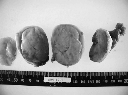
Fig. 2 A smooth-surfaced, yellowish, solid, and movable tumor was found to originate in the left ovary, measured 7 × 5 cm.

Fig. 3 A thecoma consisting of spindle cells with blunt ended nuclei and ill defined cytoplasm. The solid mass was consisted of fascicles of stromal cells, and some were vacuolated (H & E, (A) × 100, (B) × 400).

Fig. 4 Masson trichrome staining shows very little collagen deposition in focal stromal pattern (arrow) ((A) × 100, (B) × 400).
Reference
-
1. Fox H. Sex cord-stromal tumours of the ovary. J Pathol. 1985. 145:127–148.
Article2. Keeney GL. De Groot LJ, editor. Ovarian tumors with endocrine manifestations. Endocrinology. 2001. 4th ed. Philadelphia (PA): WB Saunders Company;2172.3. Stegner HE, Löning T. Endocrine-active tumors of the ovary. Pathologe. 2003. 24:314–322.4. Siekierska-Hellmann M, Sworczak K, Babińska A, Wojtylak S. Ovarian thecoma with androgenic manifestations in a postmenopausal woman. Gynecol Endocrinol. 2006. 22:405–408.
Article5. Shenker Y, Malozowski SN, Ayers J, Grekin RJ, Barkan AL. Steroid secretion by a virilizing lipoid cell ovarian tumor: origins of dehydroepiandrosterone sulfate. Obstet Gynecol. 1989. 74:502–506.6. Takemori M, Nishimura R, Hasegawa K. Ovarian thecoma with ascites and high serum levels of CA 125. Arch Gynecol Obstet. 2000. 264:42–44.
Article7. Wu L, Zhang W, Li H, Li L, Kong W, Lin L. Clinical analysis of 74 cases with ovarian thecoma. Zhonghua Fu Chan Ke Za Zhi. 2002. 37:101–103.8. Duun S. Bilateral virilizing hilus (Leydig) cell tumors of the ovary. Acta Obstet Gynecol Scand. 1994. 73:76–77.
Article9. Leedman PJ, Bierre AR, Martin FI. Virilizing nodular ovarian stromal hyperthecosis, diabetes mellitus and insulin resistance in a postmenopausal woman. Case report. Br J Obstet Gynaecol. 1989. 96:1095–1098.
Article10. O'Driscoll JB, Mamtora H, Higginson J, Pollock A, Kane J, Anderson DC. A prospective study of the prevalence of clear-cut endocrine disorders and polycystic ovaries in 350 patients presenting with hirsutism or androgenic alopecia. Clin Endocrinol (Oxf). 1994. 41:231–236.11. Loh KC, Lo JC, Zaloudek CJ, Fitzgerald PA. Occult virilizing ovarian tumours in postmenopausal women: problems in evaluation with reference to a case. Ann Acad Med Singapore. 1998. 27:712–716.12. Nokes JM, Claiborne HA Jr, Reingold WN. Thecoma with associated virilization. Am J Obstet Gynecol. 1959. 78:722–729.13. Scully RE. Fauci AS, Braunwald E, Isselbacher KJ, Wilson JD, Martin JB, Kasper DL, Hauser SL, editors. Ovarian tumors with endocrine manifestations. Harrison's principles of internal medicine. 1998. 14th ed. New York: McGraw-Hill;2113–2127.14. Takeuchi S, Ishihara N, Ohbayashi C, Itoh H, Maruo T. Stromal Leydig cell tumor of the ovary. Case report and literature review. Int J Gynecol Pathol. 1999. 18:178–182.15. Lee CH, Moon WS, Hong KE, Choi YK, Nam KH, Lee KH, et al. A case of hyperthecosis associated with virilization in the premenopausal woman. Korean J Obstet Gynecol. 1992. 35:150–156.16. Aboud E. A review of granulosa cell tumours and thecomas of the ovary. Arch Gynecol Obstet. 1997. 259:161–165.
Article17. Joe Y, Jung YH, Ro ES, Kim YP, Kwon SU. Two cases of theca cell tumor of the ovary. Korean J Obstet Gynecol. 1992. 35:451–455.18. Piver MS. Ovarian malignancies: diagnostic and therapeutic advances. 1987. Edinburgh: Churchill Livingstone;251–271.
- Full Text Links
-
- Actions
-
Cited
- CITED
-
- Close
- Share
-
- Similar articles
-
- A Case of Ovarian Low-Grade Stromal Sarcoma with Thecomatous Features: So-Called "Malignant Thecoma"
- A Case of Pseudo-Meigs' Syndrome with Elevated Serum CA 125 levels
- A case of thecoma causing precocious puberty at 6 years old age
- A case of malignant fibrothecoma of the ovary
- A Case of Juvenile Cystic Granulosa Cell Tumor of the Ovary
