J Korean Med Sci.
2006 Apr;21(2):368-370. 10.3346/jkms.2006.21.2.368.
Two Cases of Melasma with Unusual Histopathologic Findings
- Affiliations
-
- 1Department of Dermatology, Inha University School of Medicine, Incheon, Korea. jshin@inha.ac.kr
- 2Department of Dermatology, Ajou University School of Medicine, Suwon, Korea.
- KMID: 1781854
- DOI: http://doi.org/10.3346/jkms.2006.21.2.368
Abstract
- We reported two cases of clinically typical melasma presenting with unusual histopathologic findings. In one case, the epidermal melanocytes were markedly increased in number and protruded into the dermis, and in the other case, increased epidermal pigmentation as well as dermal melanocytosis were found. We suggested that the various treatment modalities of melasma should be applied depend on its histopathologic finding.
MeSH Terms
Figure
-
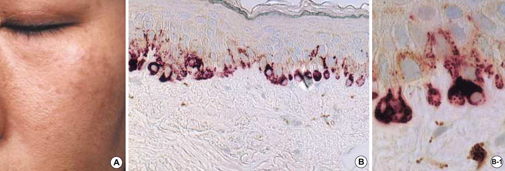
Fig. 1 (A) Malar type melasma in patient 1. (B) The melanocytes are markedly increased in number and show pendulous change (NKI-beteb, ×200), (Inset (B-1): NKI-beteb, ×1,000).
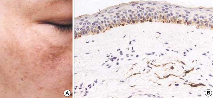
Fig. 2 (A) Malar type melasma in patient 2. (B) The lesional skin shows increased melanocytes in the epidermis and dermal dendritic pigmented cells (NKI-beteb, ×100).
Reference
-
1. Sanchez NP, Pathak MA, Sato S, Fitzpatrick TB, Sanchez JL, Mihm MC Jr. Melasma: a clinical, light microscopic, ultrastructural, and immunofluorescence study. J Am Acad Dermatol. 1981. 4:698–710.
Article2. Kang WH, Yoon KH, Lee ES, Kim J, Lee KB, Yim H, Sohn S, Im S. Melasma: histopathological characteristics in 56 Korean patients. Br J Dermatol. 2002. 146:228–237.
Article3. Kang WH, Lee ES, Choi GS. Treatment of Ota's nevus by Q-switched alexandrite laser: therapeutic outcome in relation to clinical and histopathological findings. Eur J Dermatol. 1999. 9:639–643.4. Lee ES, Kim JH, Im S, Lee KB, Sohn S, Kang WH. Application of computerized image analysis in pigmentary skin diseases. Int J Dermatol. 2001. 40:45–49.
Article5. Vennegoor C, Hageman P, Van Nouhuijs H, Ruiter DJ, Calafat J, Ringens PJ, Rumke P. A monoclonal antibody specific for cells of the melanocyte lineage. Am J Pathol. 1988. 130:179–192.6. Adema GJ, de Boer AJ, van't Hullenaar R, Denijn M, Ruiter DJ, Vogel AM, Figdor CG. Melanocyte lineage-specific antigens recognized by monoclonal antibodies NKI-beteb, HMB-50, and HMB-45 are encoded by a single cDNA. Am J Pathol. 1993. 143:1579–1585.7. Murphy GF. Elder D, Elenitsas R, Jaworsky C, Johnson B, editors. Histology of the skin. Lever's histopathology of the skin. 1997. Philadelphia: Lippincott-Raven;5–50.8. Ortonne JP, Claudy AL, Freycon F. Café au lait spots in ataxia telangiectasia (A.T.). Histochemical and ultrastructural study in one case. Arch Dermatol Res. 1980. 268:91–99.9. Bacharach-Buhles M, Lubowietzki M, Altmeyer P. Dose-dependent shift of apoptotic and unaltered melanocytes into the dermis after irradiation with UVA1. Dermatology. 1999. 198:5–10.10. Herlyn M, Berking C, Li G, Satyamoorthy K. Lessons from melanocyte development for understanding the biological events in naevus and melanoma formation. Melanoma Res. 2000. 10:303–310.
Article11. Jamal S, Schneider RJ. UV-induction of keratinocyte endothelin-1 downregulates E-cadherin in melanocytes and melanoma cells. J Clin Invest. 2002. 110:443–452.
Article
