A Case of Santorinicele without Pancreas Divisum: Diagnosis with Multi-detector Row Computed Tomography
- Affiliations
-
- 1Department of Internal Medicine, Kyung Hee University College of Medicine, Seoul, Korea. krjoo@khu.ac.kr
- KMID: 1781851
- DOI: http://doi.org/10.3346/jkms.2006.21.2.358
Abstract
- A santorinicele is defined as a focal cystic dilatation of the terminal portion of the dorsal pancreatic duct at the minor papilla. Most cases reported previously were associated with pancreas divisum and a santorinicele without pancreas divisum is known to be rare. We recently experienced a typical case of a santorinicele without pancreas divisum in a 67-yr-old woman with abdominal pain and hematochezia, subsequently proven to be the result of an ischemic colitis. The santorinicele was diagnosed incidentally with multi-detector row computed tomography using a minimum intensity projection technique, which clearly showed a cystic dilatation of the terminal portion of the dorsal pancreatic duct and a communication between the ventral and dorsal pancreatic ducts. This finding was also confirmed by a magnetic resonance cholangiopancreatography.
Keyword
MeSH Terms
Figure
-
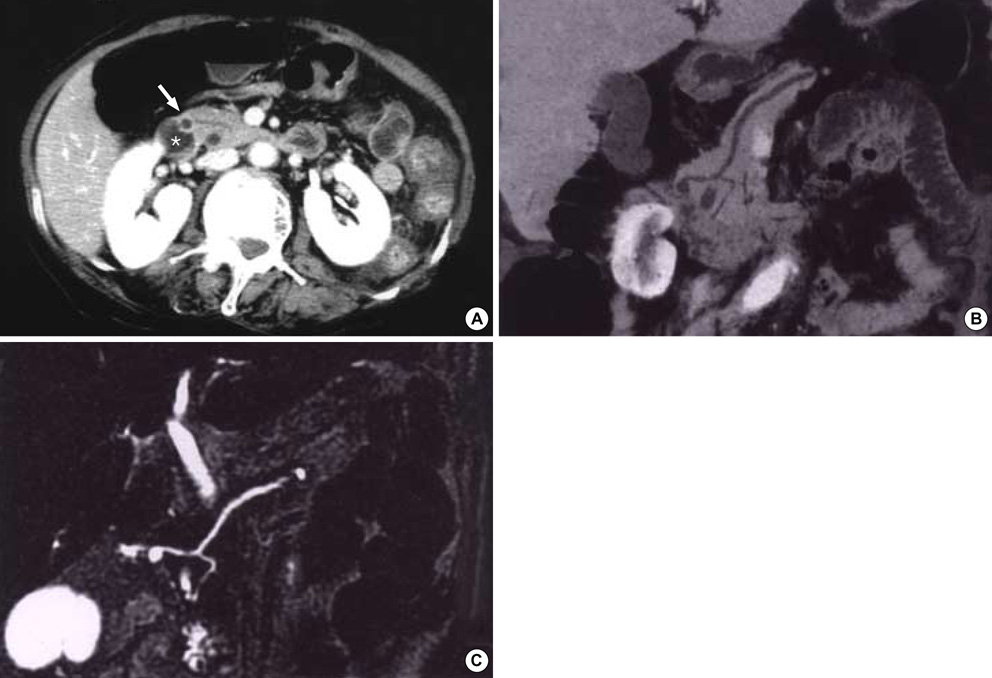
Fig. 1 (A) Contrast-enhanced axial CT scan of the abdomen at level of the pancreas head shows a small, round cystic structure (arrow) in the pancreas head adjacent to the second portion of the duodenum (asterisk). (B) CT pancreatography using a minimum intensity projection technique shows a santorinicele and communication between the ventral and dorsal pancreatic ducts. (C) Coronal MRCP image also reveals a santorinicele with no evidence of pancreas divisum.
Cited by 1 articles
-
Recurrent pancreatitis in the setting of gallbladder agenesis, ansa pancreatica, Santorinicoele and eventual intraductal papillary mucinous neoplasia (IPMN)
Sun Woo Lee, Caroline Jane Davidson, YinHiew Kia, Ben Devereaux, Savio Godinho, Mark Appleyard, Nicholas O’Rourke, Manju Dashini Chandrasegaram
Ann Hepatobiliary Pancreat Surg. 2020;24(3):381-387. doi: 10.14701/ahbps.2020.24.3.381.
Reference
-
1. Eisen G, Schutz S, Metzler D, Baillie J, Cotton PB. Santorinicele: new evidence for obstruction in pancreas divisum. Gastrointest Endosc. 1994. 40:73–76.
Article2. Peterson MS, Slivka A. Santorinicele in pancreas divisum: diagnosis with secretin-stimulated magnetic resonance pancreatography. Abdom Imaging. 2001. 26:260–263.
Article3. Costamagna G, Ingrosso M, Tringali A, Mutignani M, Manfredi R. Santorinicele and recurrent acute pancreatitis in pancreas divisum: diagnosis with dynamic secretin-stimulated magnetic resonance pancreatography and endoscopic treatment. Gastrointest Endosc. 2000. 52:262–267.
Article4. Tang H, Kay CL, Devonshire DA, Tagge E, Cotton PB. Recurrent pancreatitis in a child with pancreas divisum. Endoscopic therapy of a santorinicele. Surg Endosc. 1999. 13:1040–1043.5. Seibert DG, Matulis SR. Santorinicele as a cause of chronic pancreatic pain. Am J Gastroenterol. 1995. 90:121–123.6. Manfredi R, Costamagna G, Brizi MG, Spina S, Maresca G, Vecchioli A, Mutignani M, Marano P. Pancreas divisum and "santorinicele": diagnosis with dynamic MR cholangiopancreatography with secretin stimulation. Radiology. 2000. 217:403–408.
Article7. Joo KR, Bang SJ, Shin JW, Kim DH, Park NH. Santorinicele containing a pancreatic duct stone in a patient with incomplete pancreas divisum. Yonsei Med J. 2004. 45:952–955.
Article8. Byeon JS, Kim MH, Lee SK, Yang DH, Bae JS, Kim HJ, Lee SS, Seo DW, Min YI. Santorinicele without pancreas divisum. Gastrointest Endosc. 2003. 58:800–803.
Article
- Full Text Links
-
- Actions
-
Cited
- CITED
-
- Close
- Share
-
- Similar articles
-
- Santorinicele without Pancreas Divisum: Diagnosis with Multi-Detector Row CT and MR Cholangiopancreatography
- Three Cases of Santorinicele without Pancreas Divisum
- Santorinicele Containing a Pancreatic Duct Stone in a Patient with Incomplete Pancreas Divisum
- A Case of Successful Endoscopic Treatment for Acute Recurrent Pancreatitis Due to Pancreas Divisum with Santorinicele Masquerading as Drug Induced Pancreatitis
- A Case of Acute Pancreatitis Caused by Santorinicele with Incomplete Pancreas Divisum
