Reproducibility of Computer-Aided Detection Marks in Digital Mammography
- Affiliations
-
- 1Department of Radiology, Konkuk University Hospital, Seoul, Korea. moonwk@radcom.snu.ac.kr
- 2Department of Radiology, Seoul National University College of Medicine and Clinical Research Institute, Seoul National University Hospital and the Institute of Radiation Medicine, Seoul National University Medical Research Center, Seoul, Korea.
- 3Department of Radiology, Boramae Municipal Hospital, Seoul, Korea.
- KMID: 1779434
- DOI: http://doi.org/10.3348/kjr.2007.8.3.198
Abstract
OBJECTIVE
To evaluate the performance and reproducibility of a computer-aided detection (CAD) system in mediolateral oblique (MLO) digital mammograms taken serially, without release of breast compression. MATERIALS AND METHODS: A CAD system was applied preoperatively to the full-field digital mammograms of two MLO views taken without release of breast compression in 82 patients (age range: 33-83 years; mean age: 49 years) with previously diagnosed breast cancers. The total number of visible lesion components in 82 patients was 101: 66 masses and 35 microcalcifications. We analyzed the sensitivity and reproducibility of the CAD marks. RESULTS: he sensitivity of the CAD system for first MLO views was 71% (47/66) for masses and 80% (28/35) for microcalcifications. The sensitivity of the CAD system for second MLO views was 68% (45/66) for masses and 17% (6/35) for microcalcifications. In 84 ipsilateral serial MLO image sets (two patients had bilateral cancers), identical images, regardless of the existence of CAD marks, were obtained for 35% (29/84) and identical images with CAD marks were obtained for 29% (23/78). Identical images, regardless of the existence of CAD marks, for contralateral MLO images were 65% (52/80) and identical images with CAD marks were obtained for 28% (11/39). The reproducibility of CAD marks for the true positive masses in serial MLO views was 84% (42/50) and that for the true positive microcalcifications was 0% (0/34). CONCLUSION: The CAD system in digital mammograms showed a high sensitivity for detecting masses and microcalcifications. However, reproducibility of microcalcification marks was very low in MLO views taken serially without release of breast compression. Minute positional change and patient movement can alter the images and result in a significant effect on the algorithm utilized by the CAD for detecting microcalcifications.
MeSH Terms
Figure
-
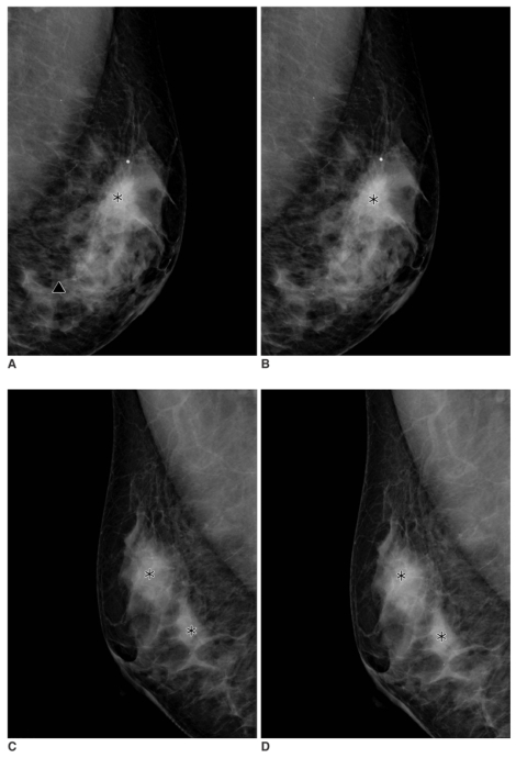
Fig. 1 A 49-year-old woman with an infiltrating ductal carcinoma in the left breast that was confirmed by a biopsy.A, B. Serial mediolateral oblique digital mammograms taken without release of breast compression within 17 seconds. The computer-aided detection system marks the mass in the left upper breast correctly on both mediolateral oblique images (asterisks). The false positive calcification mark (triangle) is seen below the mass mark on the first mediolateral oblique view, but not on the second mediolateral oblique view.C, D. Serial mediolateral oblique digital mammograms of contralateral breast taken within 17 seconds. The computer-aided detection system marks normal parenchyma areas on the first mediolateral oblique view (C) (asterisks) and the computer-aided detection system marks the false positive mass repeatedly on the second mediolateral oblique view (D).
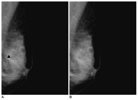
Fig. 2 A 56-year-old woman with malignant microcalcifications in the left breast.A, B. Serial mediolateral oblique digital mammograms taken without release of breast compression within 17 seconds. The computer-aided detection system marks the microcalcifications correctly on the first mediolateral oblique view (A) (triangle). The computer-aided detection system, however, does not mark the microcalcifications on the second mediolateral oblique view (B).
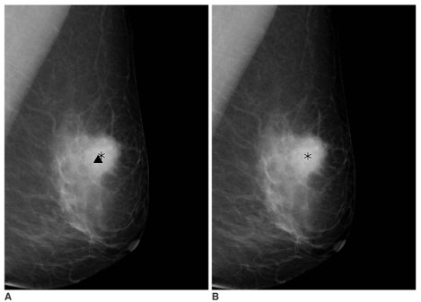
Fig. 3 A 61-year-old woman presented with a screening-detected breast cancer in the left breast.A,B. Serial mediolateral oblique digital mammograms taken without release of breast compression within 19 seconds. The computer-aided detection system marks the mass component and the microcalcifications component correctly on the first mediolateral oblique view (A) (asterisk and triangle). The computer-aided detection system, however, does not mark the microcalcifications on the second mediolateral oblique view (B).
Reference
-
1. Warren Burhenne LJ, Wood SA, D'Orsi CJ, Feig SA, Kopans DB, O'Shaughnessy KF, et al. Potential contribution of computer-aided detection to the sensitivity of screening mammography. Radiology. 2000; 215:554–562. PMID: 10796939.
Article2. Freer TW, Ulissey MJ. Screening mammography with computer-aided detection: prospective study of 12,860 patients in a community breast center. Radiology. 2001; 220:781–786. PMID: 11526282.
Article3. Zheng B, Hardesty LA, Poller WR, Sumkin JH, Golla S. Mammography with computer-aided detection: reproducibility assessment initial experience. Radiology. 2003; 228:58–62. PMID: 12759470.4. Malich A, Azhari T, Bohm T, Fleck M, Kaiser WA. Reproducibility - an important factor determining the quality of computer-aided detection (CAD) systems. Eur J Radiol. 2000; 36:170–174. PMID: 11091020.5. Taylor CG, Champness J, Reddy M, Taylor P, Potts HW, Given-Wilson R. Reproducibility of prompts in computer-aided detection (CAD) of breast cancer. Clin Radiol. 2003; 58:733–738. PMID: 12943648.
Article6. Baum F, Fischer U, Obenauer S, Grabbe E. Computer-aided detection in direct digital full-field mammography: initial results. Eur Radiol. 2002; 12:3015–3017. PMID: 12439584.
Article7. American College of Radiology. Mammography quality control manual. 1999. Reston, VA: American College of Radiology;p. 284–285.8. American College of Radiology. Breast imaging reporting and data system: BI-RADS atlas. 2003. 4th ed. Reston, VA: American College of Radiology.9. Baker JA, Lo JY, Delong DM, Floyd CE. Computer-aided detection in screening mammography: variability in cues. Radiology. 2004; 233:411–417. PMID: 15358850.
Article
- Full Text Links
-
- Actions
-
Cited
- CITED
-
- Close
- Share
-
- Similar articles
-
- Digital Mammography
- Reproducibility of Computer-Aided Detection System in Digital Mammograms
- 3D Computer-Aided Detection for Digital Breast Tomosynthesis: Comparison with 2D Computer-Aided Detection for Digital Mammography in the Detection of Calcifications
- Detection of Breast Mass in Mammogram Using Computer-Aided Diagnosis System
- Screening Mammography-Detected Cancers: The Sensitivity of the Computer-aided Detection System as Applied to Full-field Digital Mammography
