Ultrastructural Analysis of in vivo Expanded Corneal Epithelium on Amniotic Membrane
- Affiliations
-
- 1Department of Ophthalmology, Chung-Ang University College of Medicine, Seoul, Korea. Jck50ey@kornet.net
- 2Department of Pathology, Chung-Ang University College of Medicine, Seoul, Korea.
- KMID: 1778442
- DOI: http://doi.org/10.3346/jkms.2006.21.3.544
Abstract
- The purpose of this study is to characterize and compare the ultrastructural changes occurring during the in vivo cultivation of corneal epithelium on amniotic membrane (AM) at several different time points. Corneal burn patients (n=7) with a corneal epithelial defect and severe limbal damage were selected. Initially, AM transplantation with limbal autograft was performed at the acute stage of corneal burn to reconstruct the damaged ocular surface. One to six (mean interval; 3.3+/-1.2) months later, the central part of AM containing an in vivo expanded corneal epithelium was excised and retransplanted in adjacent lesions. The excised epithelium with AM was examined by electron microscopy and immunohistochemical study. By electron microscopy, one and two months after expansion, cultivated epithelium on AM showed an undifferentiated epithelium and an incomplete basement membrane (BM). But, after three months, the cultivated epithelium began to differentiate into a multilayered epithelium with a continuous BM with increased hemidesmosomes. These findings were further confirmed by immunohistochemical study, that cytokeratin K3 was expressed in the cultivated corneal epithelium and newly formed BM was partially positive of collagen IV at three months. At least 3 months may be needed for the proliferation and differentiation of in vivo cultivated corneal epithelium on AM.
MeSH Terms
Figure
-
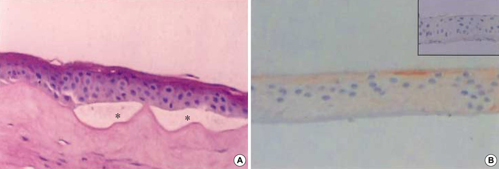
Fig. 1 First month after in vivo cultivation on amniotic membrane (AM). (A) The expanded epithelium on AM consists of 4, 5 cell layers of corneal epithelium and looks normal in appearance, though there are partial detachments (asterisk) between basal cells and AM (H&E, ×200). (B) Cultured corneal epithelium stained poorly for cytokeratin K3 (×200) and growing BM does not stain for collagen IV (inset: ×200).
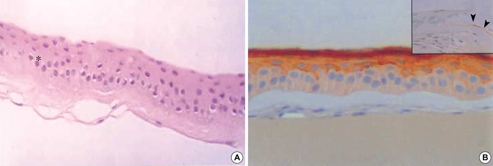
Fig. 2 Third month after in vivo cultivation on AM. (A) The cultured epithelium exhibits well differentiated five to six stratified cell layers and is attached firmly to the AM (H&E, ×200). (B) Whole suprabasal corneal epithelium is stained strongly with cytokeratin K3 (×200) and newly formed BM is stained partially with collagen IV (arrowhead in inset: ×200).

Fig. 3 EM findings of epithelial basal cells (asterisk) and hemidesmosomes (arrow) and basement membrane development. (A, B) First month (C, D) Second month (E, F) Third month. The basal cells of in vivo cultivated corneal epithelium are differentiated from rudimental round cell with large N/C ratio at the 1st month to normal cuboidal cells after 3 months of cultivation. The electron-dense structure of BM began to appear at 2 months and became distinct continuous layers with well-formed hemidesmosomes. The density of hemidesmosomes increased with time, i.e., 1.3, 2.2, and 3.4 per micrometer on average after 1, 2, 3 months of in vivo cultivation, respectively.
Cited by 2 articles
-
The Effect of In Vivo Grown Corneal Epithelium Transplantation on Persistent Epithelial Defects with Limbal Stem Cell Deficiency
Jee Taek Kim, Yeoun Sook Chun, Geo Young Song, Jae Chan Kim
J Korean Med Sci. 2008;23(3):502-508. doi: 10.3346/jkms.2008.23.3.502.Effects of Intravitreal Amniotic Membrane Injection in a Rat Model of Choroidal Neovascularization
Jong Hyun Lee, Jae Lim Chung, Samin Hong, Hyoung Jun Koh
J Korean Ophthalmol Soc. 2008;49(7):1154-1158. doi: 10.3341/jkos.2008.49.7.1154.
Reference
-
1. Schermer A, Galvin S, Sun TT. Differentiation-related expression of a major 64K corneal keratin in vivo and in culture suggests limbal location of corneal epithelial stem cells. J Cell Biol. 1986. 103:49–62.
Article2. Tseng SC, Chen JY, Huang AJ. Classification of conjunctival surgeries for corneal disease based on stem cell concept. Ophthalmic Clin North Am. 1990. 3:595–610.3. Kenyon KR, Tseng SC. Limbal autograft transplantation for ocular surface disorders. Ophthalmology. 1989. 96:709–723.
Article4. Kim JS, Kim JC, Na BK, Jeong JM, Song CY. Amniotic membrane patching promotes healing and inhibits protease activity on wound healing following acute corneal alkali burns. Exp Eye Res. 1998. 70:329–337.5. Meller D, Pires RT, Mack RJ, Figueiredo F, Heiligenhaus A, Park WC, Prabhasawat P, John T, McLeod SD, Steuhl KP, Tseng SC. Amniotic membrane transplantation for acute chemical or thermal burns. Ophthalmology. 2000. 107:980–990.
Article6. Tsubota K, Satake Y, Ohyama M, Toda I, Takano Y, Ono M, Shinozaki N, Shimazaki J. Surgical reconstruction of the ocular surface in advanced ocular cicatricial phemphigoid and Stevens-Johnson syndrome. Am J Ophthalmol. 1996. 122:38–52.7. Tseng SC, Prabhasawat P, Barton K, Gray T, Meller D. Amniotic membrane transplantation with or without limbal allografts for corneal surface reconstruction in patients with limbal stem cell deficiency. Arch Ophthalmol. 1998. 116:431–441.
Article8. Park GS, Ye J, Kim JC. Stepwise surgical approach for in vivo expansion of epithelial stem cells to treating severe acute chemical burns with total limbal deficiency. Korean J Ophthalmol. 2003. 17:75–82.9. Tsai RJ, Li LM, Chen JK. Reconstruction of damaged corneas by transplantation of autologous limbal epithelial cells. N Engl J Med. 2000. 343:86–93.
Article10. Koizumi N, Kinoshita S. Cultivated corneal epithelial stem cell transplantation in ocular surface disorder. Ophthalmology. 2001. 124:1569–1574.11. Roper-Hall MJ. Thermal and chemical burns. Trans Ophthalmol Soc UK. 1965. 85:631–653.12. Lee SH, Tseng SC. Amniotic membrane transplantation for persistent epithelial defects with ulceration. Am J Ophthalmol. 1997. 123:303–312.
Article13. Tisdale AS, Spurr-Michaud SJ, Rodrigues M, Hackett J, Krachmer J, Gipson IK. Development of the anchoring structures of the epithelium in rabbit and human fetal corneas. Invest Ophthalmol Vis Sci. 1988. 29:727–736.14. Kim JC, Kim YJ, Song HJ. Down-regulation of metalloproteinases and inducible nitiric oxide synthese in keratocyte cultured with amniotic membrane extract (ARVO abstract no 1393). Invest Ophthalmol Vis Sci. 2000. 41:S265.15. Na BK, Hwang JH, Kim JC. Analysis of human amniotic membrane components as proteinase inhibitors for development of therapeutic agent for recalcitrant keratitis. Trophoblast Res. 1999. 13:453–466.
Article16. Kim JC, Tseng SC. Transplantation of preserved human amniotic membrane for surface reconstruction in severely damaged rabbit corneas. Cornea. 1995. 14:473–484.
Article17. Kim JC, Tseng SC. The effects on inhibition of corneal neovascularization after human amniotic membrane transplantation in severely damaged rabbit corneas. Korean J Ophthalmol. 1995. 9:32–46.
Article18. Tseng SC, Tsubota K. Important concepts for treating ocular surface and tear disorders. Am J Ophthalmol. 1997. 124:825–835.
Article19. Boudreau N, Sympson CJ, Werb Z, Bissell MJ. Suppression of ICE and apoptosis in mammary epithelial cells by extracellular matrix. Science. 1995. 267:891–893.
Article20. Meller D, Pires RT, Tseng SC. Ex-vivo preservation and expansion of human limbal epithelial progenitor cells by amniotic membrane. Invest Ophthalmol Vis Sci. 1999. 40:S329.21. Azuara-Blanco A, Pillai CT, Dua HS. Amniotic membrane transplantation for ocular surface reconstruction. Br J Ophthalmol. 1999. 83:399–402.
Article22. Letko E, Stechschulte SU, Kenyon KR, Sadeq N, Romero TR, Samson CM, Nguyen QD, Harper SL, Primack JD, Azar DT, Gruterich M, Dohlman CH, Baltatzis S, Foster CS. Amniotic membrane inlay and overlay grafting for corneal epithelial defects and stromal ulcers. Arch Ophthalmol. 2001. 119:659–663.
Article
- Full Text Links
-
- Actions
-
Cited
- CITED
-
- Close
- Share
-
- Similar articles
-
- Morphology and Adhesion Complex of Cultured Epithelium, on Amniotic Membrane in Vitro and in Vivo
- New Strategy of Ocular Surface Disease: Ocular Surface Reconstruction Using Amniotic Membrane and Limbal Stem Cell Transplantation
- Transplantation of in vivo Cultivated Limbal Corneal Epithelial Cells with Total Limbal Stem Cell Deficiency
- Corneal Tattooing to Mask the Opacification after Amniotic Membrane Grafting for Corneal Ulcer
- Effect of Amniotic Membrane on Epithelial Thickness and Formation of Hemidesmosomes after Corneal Stromal Wound
