J Korean Med Sci.
2006 Jun;21(3):474-477. 10.3346/jkms.2006.21.3.474.
Incontinentia Pigmenti: Clinical Observation of 40 Korean Cases
- Affiliations
-
- 1Department of Dermatology, Seoul National University College of Medicine, Seoul, Korea. oskwon@snu.ac.kr
- 2Department of Dermatology, College of Medicine, Chung-Ang University, Seoul, Korea.
- KMID: 1778431
- DOI: http://doi.org/10.3346/jkms.2006.21.3.474
Abstract
- Incontinentia pigmenti (IP) is an uncommon genodermatosis that usually occurs in female infants. It is characterized by ectodermal, mesodermal, neurological, ocular, and dental manifestations. The aim of this study was to clarify clinical symptoms, accompanying diseases, and complications of IP. Forty cases of IP have been reviewed by their medical records, laboratory data, clinical photographs, and telephone survey. Male-to-female ratio was 1 to 19 and their onsets were mostly in utero. They were usually diagnosed during the neonatal period owing to their early expression of skin manifestation. Central nervous system anomalies were found in 46.7%. Ocular disorders and dental defects were detected in 66.7% and 72.7% respectively. The most commonly diagnosed anomalies were hypodontia, retinopathy, and seizure. For better understanding of IP, long term and close cooperation between dermatologists, pediatricians, neuroscientists, genentic counselors, and even dentists is crucial.
MeSH Terms
-
Stomatognathic Diseases/complications
Skin Diseases/complications
Male
Magnetic Resonance Imaging/methods
Korea
Infant, Newborn
Infant
Incontinentia Pigmenti/*diagnosis/pathology
Humans
Female
Eye Diseases/complications
Eosinophilia/complications
Child, Preschool
Child
Central Nervous System Diseases/complications
Figure
-
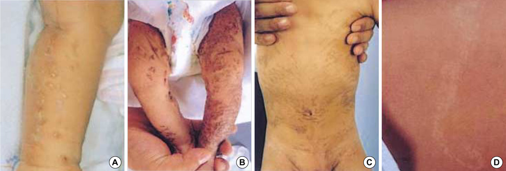
Fig. 1 Clinical stages of incontinentia pigmenti. (A) Linear vesicles and bullae of stage 1. (B) Dark brown colored verrucous papules and plaques of stage 2. (C) Light brown colored swirling hyperpigmentation of stage 3. (D) White atrophic patches of stage 4.
Cited by 1 articles
-
The Common NF-κB Essential Modulator (NEMO) Gene Rearrangement in Korean Patients with Incontinentia Pigmenti
Min-Jung Song, Jong-Hee Chae, Eun-Ae Park, Chang-Seok Ki
J Korean Med Sci. 2010;25(10):1513-1517. doi: 10.3346/jkms.2010.25.10.1513.
Reference
-
1. Berlin AL, Paller AS, Chan LS. Incontinentia pigmenti: a review and update on the molecular basis of pathophysiology. J Am Acad Dermatol. 2002. 47:169–187.
Article2. Cohen BA. Incontinentia pigmenti. Neurol Clin. 1987. 5:361–377.3. Landy SJ, Donnai D. Incontinentia pigmenti (Bloch-Sulzberger syndrome). J Med Genet. 1993. 30:53–59.
Article4. Macey-Dare LV, Goodman JR. Incontinentia pigmenti: seven cases with dental manifestations. Int J Paediatr Dent. 1999. 9:293–297.
Article5. Carney RG. Incontinentia pigmenti. A world statistical analysis. Arch Dermatol. 1976. 112:535–542.
Article6. Kenwrick S, Woffendin H, Jakins T, Shuttleworth SG, Mayer E, Greenhalgh L, Whittaker J, Rugolotto S, Bardaro T, Esposito T, D'Urso M, Soli F, Turco A, Smahi A, Hamel-Teillac D, Lyonnet S, Bonnefont JP, Munnich A, Aradhya S, Kashork CD, Shaffer LG, Nelson DL, Levy M, Lewis RA. International IP Consortium. Survival of male patients with incontinentia pigmenti carrying a lethal mutation can be explained by somatic mosaicism or Klinefelter syndrome. Am J Hum Genet. 2001. 69:1210–1217.7. Scheuerle AE. Male cases of incontinentia pigmenti: case report and review. Am J Med Genet. 1998. 77:201–218.
Article8. Moss C. Cytogenetic and molecular evidence for cutaneous mosaicism: the ectodermal origin of Blaschko lines. Am J Med Genet. 1999. 85:330–333.
Article9. Cohen PR. Incontinentia pigmenti: clinicopathologic characteristics and differential diagnosis. Cutis. 1994. 54:161–166.10. Hadj-Rabia S, Froidevaux D, Bodak N, Hamel-Teilloc D, Smahi A, Touil Y, Fraitag S, de Prost Y, Bodemer C. Clinical study of 40 cases of incontinentia pigmenti. Arch Dermatol. 2003. 139:1163–1170.
Article11. Phan TA, Wargon O, Turner AM. Incontinentia pigmenti case series: clinical spectrum of incontinentia pigmenti in 53 female patients and their relatives. Clin Exp Dermatol. 2005. 30:474–480.
Article12. el-Benhawi MO, George WM. Incontinentia pigmenti. Cutis. 1988. 41:259–262.13. Berretty PJ, Cormane RH. Eosinophilic granulocytes and skin disorders. Int J Dermatol. 1981. 20:531–540.
Article14. Chun IK, Chung TB, Kim YP. Clinical observation of incontinentia pigmenti. Korean J Dermatol. 1985. 23:171–176.15. Patrizi A, Neri I, Guareschi E, Cocchi G. Bullous recurrent eruption of incontinentia pigmenti. Pediatr Dermatol. 2004. 21:613–614.
Article16. Wiederholt T, Poblete-Gutierrez P, Ott H, Lehmann S, Grussendorf-Conen EI, Beermann T, Frank J. Incontinentia pigmenti in a five-week-old girl. Hautarzt. 2004. 55:999–1001.
