Obstructive Jaundice Caused by Clonorchiasis-associated Duodenal Papillitis: A Case Report
- Affiliations
-
- 1Department of Internal Medicine, East-West Neo Medical Center, Kyung Hee University School of Medicine, Seoul, Korea. krjoo@khu.ac.kr
- 2Department of Pathology, East-West Neo Medical Center, Kyung Hee University School of Medicine, Seoul, Korea.
- KMID: 1777988
- DOI: http://doi.org/10.3346/jkms.2011.26.1.135
Abstract
- We describe an unusual presentation of Clonorchis sinensis infection with obstructive jaundice due to duodenal papillitis which was relieved dramatically by endoscopic sphincterotomy. A 26-yr-old male presented with complaints of fatigue, weight loss and painless jaundice. The history was significant for frequent ingestion of raw freshwater fish. The patient underwent endoscopic retrograde cholangiopancreatography for evaluation of obstructive jaundice. The duodenal papilla was markedly edematous with a bulging configuration and hyperemic changes at the orifice. Cholangiography revealed mild bile duct dilatation and irregular wall changes with multiple indentations. However, there were no biliary stricture or stones noted as the cause of obstructive jaundice. We performed an endoscopic sphincterotomy for effective bile drainage through the duodenal papilla. After the sphincterotomy, the patient's jaundice was dramatically improved. Pathology of the duodenal papilla showed eosinophilic infiltration of the mucosa. Parasitic eggs, consistent with the diagnosis of C. sinensis, were found in the bile sample.
MeSH Terms
-
Adult
*Ampulla of Vater
Animals
Anthelmintics/therapeutic use
Bile/parasitology
Cholangiopancreatography, Endoscopic Retrograde
Cholangitis/*diagnosis/parasitology/pathology
Clonorchiasis/*diagnosis
Clonorchis sinensis/drug effects/isolation & purification
Duodenum/pathology
Humans
Jaundice, Obstructive/*diagnosis/etiology
Male
Praziquantel/therapeutic use
Sphincterotomy, Endoscopic
Tomography, X-Ray Computed
Figure
-
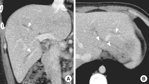
Fig. 1 CT findings of the abdomen. (A) A coronal image shows mild dilated intrahepatic bile duct of the right hepatic lobe (arrows). (B) An axial image shows dilated intrahepatic bile duct of the left hepatic lobe (arrowheads).
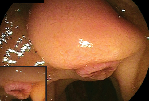
Fig. 2 Duodenoscopy shows edematous and bulging duodenal papilla with hyperemic orifice.
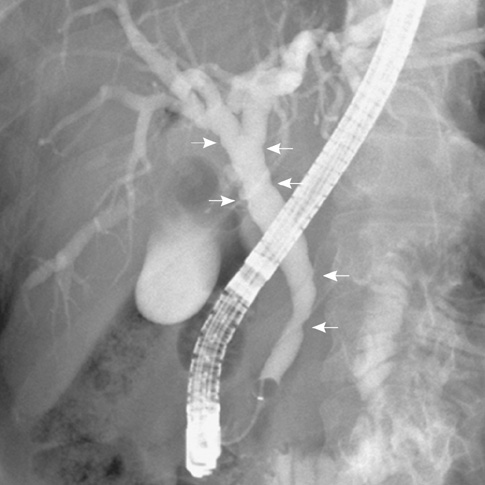
Fig. 3 Endoscopic retrograde cholangiography shows mild bile duct dilatation with multiple ductal irregularities and indentations (arrows).
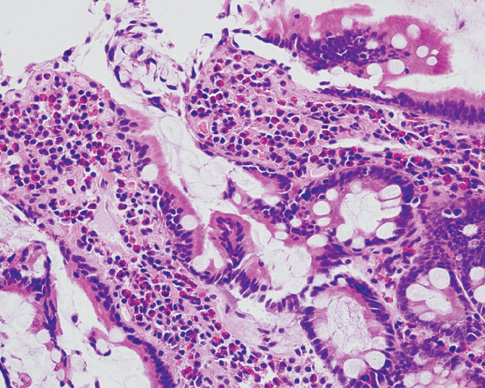
Fig. 4 Pathological findings of the biopsy specimen show many eosinophilic cells in the lamina propria of the mucosal layer (H&E, × 400).
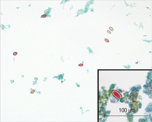
Fig. 5 Eggs of C. sinensis are found in the bile fluid sample (Pap, × 200), inset, × 400.
Cited by 1 articles
-
Cytochrome C Oxidase Subunit 1, Internal Transcribed Spacer 1, Nicotinamide Adenine Dinucleotide Hydrogen Dehydrogenase Subunits 2 and 5 of Clonorchis sinensis Ancient DNA Retrieved from Joseon Dynasty Mummy Specimens
Jong Ha Hong, Chang Seok Oh, Jong-Yil Chai, Min Seo, Dong Hoon Shin
J Korean Med Sci. 2019;34(20):. doi: 10.3346/jkms.2019.34.e149.
Reference
-
1. Rim HJ. Clonorchiasis: an update. J Helminthol. 2005. 79:269–281.2. Kim EM, Kim JL, Choi SY, Kim JW, Kim S, Choi MH, Bae YM, Lee SH, Hong ST. Infection status of freshwater fish with metacercariae of clonorchis sinensis in Korea. Korean J Parasitol. 2008. 46:247–251.3. Cho SH, Lee KY, Lee BC, Cho PY, Cheun HI, Hong ST, Sohn WM, Kim TS. Prevalence of clonorchiasis in southern endemic areas of Korea in 2006. Korean J Parasitol. 2008. 46:133–137.4. Stunell H, Buckley O, Geoghegan T, Torreggiani WC. Recurrent pyogenic cholangitis due to chronic infestation with Clonorchis sinensis (2006: 8b). Eur Radiol. 2006. 16:2612–2614.5. Sun T. Clonorchiasis: a report of four cases and discussion of unusual manifestations. Am J Trop Med Hyg. 1980. 29:1223–1227.6. Sullivan WG, Koep LJ. Common bile duct obstruction and cholangiohepatitis in clonorchiasis. JAMA. 1980. 243:2060–2061.7. Choi D, Hong ST. Imaging diagnosis of clonorchiasis. Korean J Parasitol. 2007. 45:77–85.8. Choi TK, Wong KP, Wong J. Cholangiographic appearance in clonorchiasis. Br J Radiol. 1984. 57:681–684.9. Park JS, Kim MH, Lee SK, Seo DW, Lee SS, Chang HS, Han J, Kim JS, Min YI. The clinical significance of papillitis of the major duodenal papilla. Gastrointest Endosc. 2002. 55:877–882.10. Lai CH, Chin C, Chung HC, Liu H, Hwang JC, Lin HH. Clonorchiasis-associated perforated eosinophilic cholecystitis. Am J Trop Med Hyg. 2007. 76:396–398.11. Shen C, Kim JH, Lee JK, Bae YM, Choi MH, Oh JK, Lim MK, Shin HR, Hong ST. Collection of clonorchis sinensis adult worms from infected humans after praziquantel treatment. Korean J Parasitol. 2007. 45:149–152.
- Full Text Links
-
- Actions
-
Cited
- CITED
-
- Close
- Share
-
- Similar articles
-
- The effects of endoscopic shincteropapillotomy in clonorchiasis patients with obstructive jaundice
- A Case of Obstructive Jaundice after Insertion of Metallic Stent for Duodenal Obstruction by Recurrent Duodenal Ulcer
- A Case of Anomalous Drainage of the Common Bile Duct into the Duodenal Bulb Presenting with Obstructive Jaundice
- ERCP Findings in Clonorchiasis
- A Case of Obstructive Jaundice due to Tuberculous Lymphadenitis with Duodenal Tuberculosis
