Unusual Cause of Acute Right Ventricular Dysfunction: Rapid Progression of Superior Vena Cava Aneurysm Complicated by Thrombosis and Pulmonary Thromboembolism
- Affiliations
-
- 1Department of Cardiothoracic Surgery, Chonnam National University Hospital, Gwangju, Korea.
- 2Department of Cardiovascular Medicine, Chonnam National University Hospital, Gwangju, Korea. christiankyehun@hanmail.net
- 3Department of Radiology, Chonnam National University Hospital, Gwangju, Korea.
- KMID: 1777872
- DOI: http://doi.org/10.3346/jkms.2011.26.5.690
Abstract
- Aneurysms of the major thoracic veins are rare. They are usually asymptomatic and thus treated conservatively. We report an extremely rare case of rapidly progressing superior vena cava (SVC) aneurysm complicated by thrombosis and acute pulmonary thromboembolism (PTE) with right ventricular dysfunction. Thrombolytic therapy for hemodynamically significant acute PTE was harmful to the patient in the present case, because it induced further thrombosis and mobilization of the thrombi within the aneurysm, subsequently causing de novo PTE. Surgical aneurysmectomy combined with pulmonary artery embolectomy would be a treatment of choice in patients with SVC aneurysm complicated by acute PTE.
Keyword
MeSH Terms
Figure
-

Fig. 1 Serial radiographic changes of the superior vena cava (SVC) aneurysm. Chest radiography at 2 yr ago (A) and 1 month (B) ago revealed the SVC aneurysm without any changes in size. The SVC aneurysm was markedly increased in size on chest radiography at admission (C) and disappeared after surgery (D).

Fig. 2 Multiplanar reformat images (A, B) and three-dimensional volume rendered images (C, D) of chest computed tomographic angiography demonstrated the superior vena cava (SVC) aneurysm with internal thrombi (★) protruding into the SVC through the narrow neck (arrow head in A and D) and pulmonary arterial embolism (arrow heads in B).
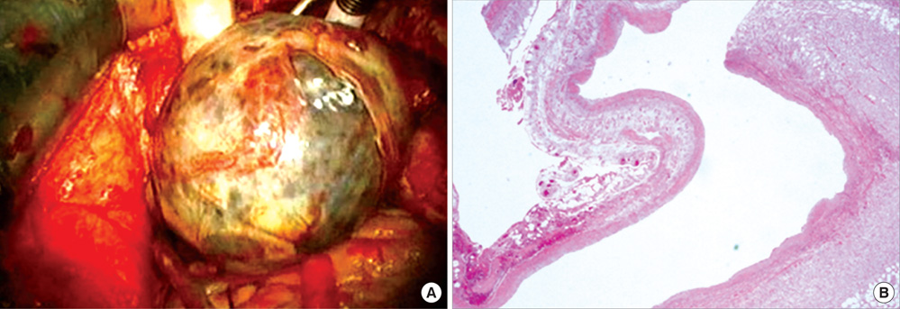
Fig. 3 A giant saccular aneurysm with internal thrombi formation arising from the superior vena cava was shown on surgical fields (A). On histopathologic examination, the aneurysmal wall contained all three layers of vascular wall, indicating true aneurysm (B).
Reference
-
1. Schatz IJ, Fine G. Venous aneurysms. N Engl J Med. 1962. 266:1310–1312.2. Rappaport DC, Ros PR, Moser RP Jr. Idiopathic dilatation of the thoracic venous system. Can Assoc Radiol J. 1992. 43:385–387.3. Koga S, Ikeda S, Sanuki Y, Ninomiya A, Izumikawa T, Miyahara Y, Kohno S. A case of asymptomatic fusiform aneurysm of the superior vena cava detected by magnetic resonance imaging. Int J Cardiol. 2006. 113:e39–e41.4. Taira A, Akita H. Ruptured venous aneurysm of the persistent left superior vena cava. Angiology. 1981. 32:656–659.5. Pasic M, Schöpke W, Vogt P, von Segesser L, Schneider J, Turina M. Aneurysm of the superior mediastinal veins. J Vasc Surg. 1995. 21:505–509.6. Gomez MA, Delhommais A, Presicci PF, Besson M, Roger R, Alison D. Partial thrombosis of an idiopathic azygos vein aneurysm. Br J Radiol. 2004. 77:342–343.7. Ream CR, Giardina A. Congenital superior vena cava aneurysm with complications caused by infectious mononucleosis. Chest. 1972. 62:755–757.8. Nakamura Y, Nakano K, Nakatani H, Fukuda T, Honda K, Homma N. Surgical exclusion of a thrombosed azygos vein aneurysm causing pulmonary embolism. J Thorac Cardiovasc Surg. 2007. 133:834–835.
- Full Text Links
-
- Actions
-
Cited
- CITED
-
- Close
- Share
-
- Similar articles
-
- Pseudoaneurysm of Surgically Reconstructed Right Ventricular Outflow Tract Complicated by Superior Vena Cava Syndrome
- A Case of Behcet's Disease with Superior Vena Cava Syndrome
- Superior Vena Cava Thrombosis Treated Successfully by Percutaneous Insertion of Metallic Stent in a Patient with Behcet's Disease
- lilac Vein Thrombosis: A Case Report of Treatment with Inferior Vena Cava Filter, Urokinase and Vascular Stent
- Retrograde Tempofilter II(TM) Placement within the Superior Vena Cava in a Patient with Acute Upper Extremity Deep Venous Thrombosis: the Filter Stands on Its Head
