Pulmonary Metastases of Alveolar Soft-Part Sarcoma: CT Findings in Three Patients
- Affiliations
-
- 1Department of Radiology, Seoul National University College of Medicine, Seoul, Korea.
- KMID: 1777297
- DOI: http://doi.org/10.3348/kjr.2000.1.1.56
Abstract
- Alveolar soft-part sarcoma is a rare soft tissue sarcoma of young adults with unknown histogenesis, and the organ most frequently involved in metastasis is the lung. We report the CT findings of three patients of pulmonary metastases of alveolar soft-part sarcoma, which manifested as clearly enhanced pulmonary nodules or masses. On enhanced scans, some of the masses were seen to contain dilated and tortuous intratumoral vessels.
MeSH Terms
Figure
-
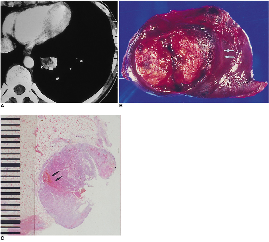
Fig. 1 A-32-year-old woman with a history of excision of alveolar soft part sarcoma involving the left foot 5 years earlier. A. Enhanced CT scan shows a well-defined, enhanced nodule. Clearly enhanced tubular structures are seen within the lesion (arrowheads), suggesting dilated vessels (attenuation: 9.2±8 H). B. Resected lung specimen shows a 2×1.5×1.5 cm sized nodule surrounded by pseudocapsule (arrows). C. Light microscopic examination demonstrates a clearly circunscribed nodule containing dilated vascular structures (arrows) which correspond to the enhanced tubular structures seen in A. The ruler in the right side of the picture is graduated in millimeters.

Fig. 2 A-31-year-old woman who presented with mass in the left leg, cough, and blood tinged sputum. Enhanced CT scan reveals multiple metastatic nodules in both lungs, with variable degrees of enhancement. Note the presence of an enhanced tubular vascular structure within a mass in the right lung (arrowheads).
Cited by 1 articles
-
Pulmonary Metastasis Originated from Uterine Sarcoma, Presenting as Multiple Nodules with Tortuous, Serpentine, Aneurysmal, Dilated Intratumoral Vessels: A Case Report
Hyeon Ji Jang, Song Soo Kim, Hee Sun Park, Jeong Eun Lee, Jin Hwan Kim
J Korean Soc Radiol. 2018;78(4):284-288. doi: 10.3348/jksr.2018.78.4.284.
Reference
-
1. Radin DR, Ralls PW, Boswell WD, Lundell C, Hallas JM. Alveolar soft tissue sarcoma: CT findings. J Comput Assist Tomogr. 1984. 8:344–345.2. Lorigan JG, O'Keeffe FN, Evans HL, Wallace S. The radiologic manifestations of alveolar soft-part sarcoma. AJR. 1989. 153:335–339.3. Lieberman PH, Brennan MF, Kimmel M, Erlandson RA, Garin-Chesa P, Flehinger BY. Alveolar soft-part sarcoma: a clinicopathologic study of half a century. Cancer. 1989. 63:1–13.4. Cordier JF, Bailly C, Tabone E, Cheix F, Brune J, Touraine R. Alveolar soft-part sarcoma presenting as asymptomatic pulmonary nodules: report of a case with ultrastructural diagnosis. Thorax. 1985. 40:203–204.5. Daly B, Cheung H, Gaines A, Bradley MJ, Metreweli C. Imaging of alveolar soft-part sarcoma. Clin Radiol. 1992. 46:253–256.6. Christopherson WM, Foote FW Jr, Stewart FW. Alveolar soft-part sarcoma: structurally characteristic tumors of uncertain histogenesis. Cancer. 1952. 5:100–111.7. Hurt R, Bates M, Harrison W. Alveolar soft-part sarcoma. Thorax. 1982. 37:877–886.8. Kodama K, Doi O, Higashiyama M, et al. Surgery for multiple metastases from alveolar soft-part sarcoma. Surg Today. 1997. 27:806–811.
- Full Text Links
-
- Actions
-
Cited
- CITED
-
- Close
- Share
-
- Similar articles
-
- Alveolar soft part Sarcoma with Metastasis to Bone: A Case Report
- Alveolar Soft-Part Sarcoma of the Female Genital Tract
- Intracerebral Metastasis of Alveolar Soft Part Sarcoma: A case report and study on its histogenesis
- Cerebral Metastases and Menifestation of the Alveolar Soft Part Sarcoma
- A Case of Alveolar Soft-Part Sarcoma
