Synchronous Double Primary Hepatic Cancer: Hepatocellular Carcinoma and Intrahepatic Cholangiocarcinoma
- Affiliations
-
- 1Department of Internal Medicine, Hanyang University College of Medicine, Seoul, Korea. noshin@hanyang.ac.kr
- 2Department of Pathology, Hanyang University College of Medicine, Seoul, Korea.
- KMID: 1775753
- DOI: http://doi.org/10.4166/kjg.2013.62.2.135
Abstract
- No abstract available.
MeSH Terms
-
Bile Duct Neoplasms/*diagnosis/pathology/radiography
Carcinoma, Hepatocellular/*diagnosis/radiography/therapy
Chemoembolization, Therapeutic
Cholangiocarcinoma/*diagnosis/pathology/radiography
Humans
Immunohistochemistry
Keratin-7/metabolism
Liver Neoplasms/*diagnosis/pathology/radiography/therapy
Male
Middle Aged
Neoplasms, Multiple Primary/*diagnosis/pathology/radiography
Positron-Emission Tomography
Tomography, X-Ray Computed
Keratin-7
Figure
-

Fig. 1. Abdominal CT scan and liver MRI findings of right (arrows) and left (arrowheads) lobe tumors. (A) Arterial phase abdominal CT scan shows 11.6 cm sized hypoattenuated mass with irregular margin in the right lobe and 4.2 cm sized early enhancing mass in the left lobe. (B) Delayed phase abdominal CT scan shows peripheral enhancement of the mass in the right lobe with central “filling in” whereas the tumor in the left lobe shows early wash out pattern. (C) Unenhanced T1-weighted liver MRI shows a hypointense mass in the right lobe and an isointense tumor in the left lobe. (D) Unenhanced T2-weight-ed liver MRI demonstrates that the tumor in the right lobe shows heterogeneous signal intensity and the tumor in the left lobe shows high signal intensity.

Fig. 2. PET-CT findings of hepatic tumors. (A) PET and (B) PET-CT show about 7–8 cm sized large mass with hypermetablic rim in the right lobe (SUVmax 6.65; arrow) and isometa-bolic mass in the left lobe (arrow head).

Fig. 3. Microscopic findings of hepatic tumors. (A) The mass in the right lobe demonstrates invasive duct-like structure lined by atypical cuboidal cells consistent with cholangiocarcinoma (H&E, ×200). (B) The mass in the left lobe demonstrates thickened hepatocytic plate with trabecular growth pattern consistent with hepatocellular carcinoma (H&E, ×200). (C) Immunohistochemical stain for cytokeratin 7 of the right hepatic mass shows immunoreactivity in the cytoplasm of tumor cells (×200). (D) Immunohistochemical stain for HepPar-1 of the left hepatic mass reveals positive staining in the cytoplasm of tumor cells (×200).
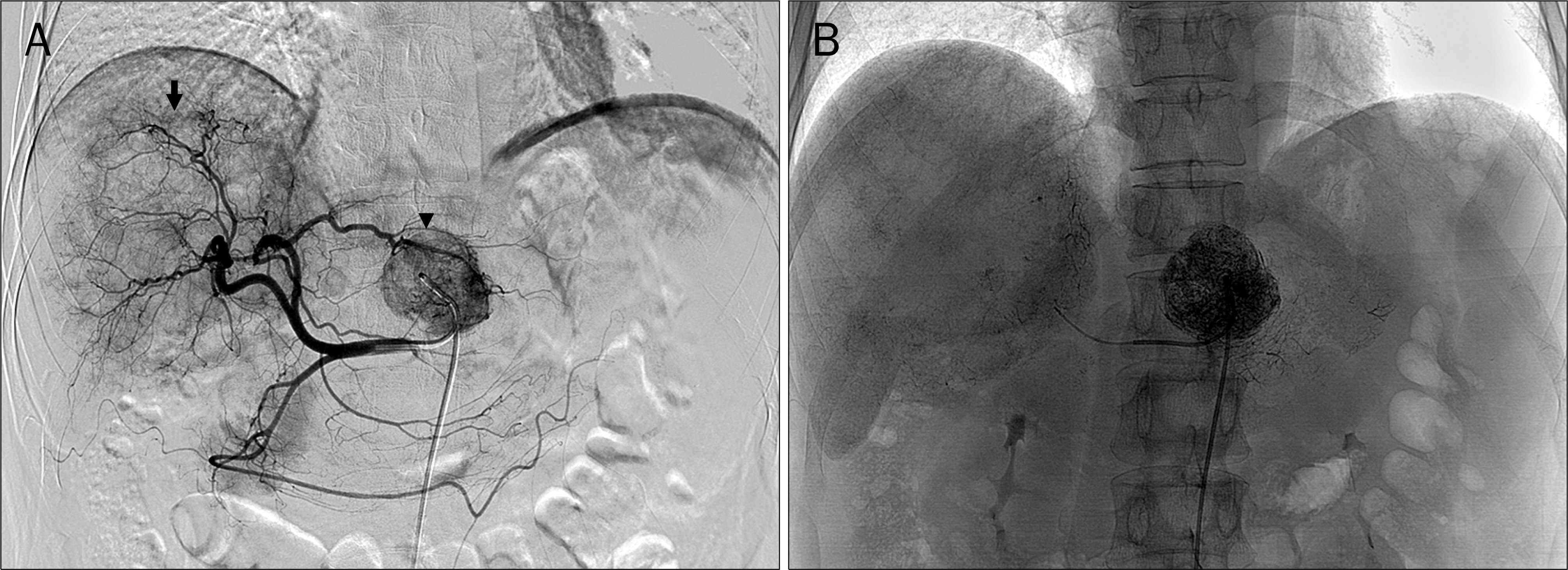
Fig. 4. Hepatic arteriography and transcatheter arterial chemoembolization (TACE) findings. (A) Hepatic arteriogram reveals huge irregular marginated mass with faint inhomogeneous tumor staining in the right lobe (arrow) and about 3.5 cm sized mass with dense tumor staining in the left lobe (arrowhead). (B) TACE was performed on the left lobe tumor.
Reference
-
References
1. Yano Y, Yamamoto J, Kosuge T, et al. Combined hepatocellular and cholangiocarcinoma: a clinicopathologic study of 26 resected cases. Jpn J Clin Oncol. 2003; 33:283–287.
Article2. Allen RA, Lisa JR. Combined liver cell and bile duct carcinoma. Am J Pathol. 1949; 25:647–655.3. Inaba K, Suzuki S, Sakaguchi T, et al. Double primary liver cancer (intrahepatic cholangiocarcinoma and hepatocellular carcinoma) in a patient with hepatitis C virus-related cirrhosis. J Hepatobiliary Pancreat Surg. 2007; 14:204–209.
Article4. Ohwada S, Yoshihiro O, Iwazaki S, et al. Double cancer in different hepatic lobes: hepatocellular and cholangiocellular carcinoma. Hepatogastroenterology. 1995; 42:411–414.5. Watanabe T, Sakata J, Ishikawa T, et al. Synchronous development of HCC and CCC in the same subsegment of the liver in a patient with type C liver cirrhosis. World J Hepatol. 2009; 1:103–109.
Article6. Fuji N, Taniguchi H, Amaike H, et al. Synchronously resected double primary hepatic cancer, hepatocellular carcinoma and cholangiocarcinoma. J Gastroenterol Hepatol. 2005; 20:967–969.
Article7. Matsuda M, Hara M, Suzuki T, Kono H, Fujii H. Synchronously resected double primary hepatic cancers – hepatocellular carcinoma and cholangiolocellular carcinoma. J Hepatobiliary Pancreat Surg. 2006; 13:571–576.
Article8. Sanada Y, Shiozaki S, Aoki H, Takakura N, Yoshida K, Yamaguchi Y. A clinical study of 11 cases of combined hepatocellular-cholangiocarcinoma Assessment of enhancement patterns on dynamics computed tomography before resection. Hepatol Res. 2005; 32:185–195.9. Park SY, Kim HS, Hong EK, Kim WH. Expression of cytokeratins 7 and 20 in primary carcinomas of the stomach and colorectum and their value in the differential diagnosis of metastatic carcinomas to the ovary. Hum Pathol. 2002; 33:1078–1085.
Article10. Kang DB, Kim SH, Byun SJ, et al. Metastatic small bowel perforation caused by intrahepatic cholangiocarcinoma in a patient with combined hepatocellular-cholangiocarcinoma. J Korean Surg Soc. 2009; 77:138–142.
Article11. Choi JH. Combined hepatocellular-cholangiocarcinoma: recent progress in pathology and classification. Yeungnam Univ J Med. 2011; 28:1–12.
Article12. Yin X, Zhang BH, Qiu SJ, et al. Combined hepatocellular carcinoma and cholangiocarcinoma: clinical features, treatment modalities, and prognosis. Ann Surg Oncol. 2012; 19:2869–2876.
Article13. Jarnagin WR, Weber S, Tickoo SK, et al. Combined hepatocellular and cholangiocarcinoma: demographic, clinical, and prognostic factors. Cancer. 2002; 94:2040–2046.14. Koh KC, Lee H, Choi MS, et al. Clinicopathologic features and prognosis of combined hepatocellular cholangiocarcinoma. Am J Surg. 2005; 189:120–125.
Article15. Lee CH, Hsieh SY, Chang CJ, Lin YJ. Comparison of clinical characteristics of combined hepatocellular-cholangiocarcinoma and other primary liver cancers. J Gastroenterol Hepatol. 2013; 28:122–127.
Article16. Singh S, Chakraborty S, Bonthu N, Radio S, Hussain SM, Sasson A. Combined hepatocellular cholangiocarcinoma: a case report and review of literature. Dig Dis Sci. 2013; 58:2114–2123.
Article17. Chi M, Mikhitarian K, Shi C, Goff LW. Management of combined hepatocellular-cholangiocarcinoma: a case report and literature review. Gastrointest Cancer Res. 2012; 5:199–202.
- Full Text Links
-
- Actions
-
Cited
- CITED
-
- Close
- Share
-
- Similar articles
-
- A Case Report of Synchronous Double Primary Liver Cancers Combined with Early Gastric Cancer
- A case with intrahepatic double cancer: hepatocellular carcinoma and cholangiocarcinoma associated with multiple von Meyenburg complexes
- Intermediate hepatic carcinoma mimicking intrahepatic cholangiocarcinoma: A case report
- Cancer Stem Cells in Primary Liver Cancers: Pathological Concepts and Imaging Findings
- Combined Hepatocellular-cholangiocarcinoma
