Spontaneous Retroperitoneal Hemorrhage and Hemothorax After Intravenous Heparin Treatment
- Affiliations
-
- 1Division of Cardiology, Department of Internal Medicine, Cheongju St. Mary's Hospital, Cheongju, Korea.
- 2Division of Cardiology, Department of Internal Medicine, Soonchunhyang University Cheonan Hospital, Cheonan, Korea. drlsj@paran.com
- KMID: 1769511
- DOI: http://doi.org/10.4070/kcj.2009.39.1.32
Abstract
- Spontaneous retroperitoneal hemorrhage is a rare complication after percutaneous coronary intervention (PCI). The patient can be in danger if bleeding is not stopped immediately. However, it is not easy to control the bleeding completely because the bleeding foci can be multiple and there is a rich network of collateral circulation. We report a case of spontaneous retroperitoneal hemorrhage successfully treated using multiple microcoils. One year later, panhypopituitarism occurred as a likely consequence of the accompanying hypovolemic shock.
Keyword
MeSH Terms
Figure
-
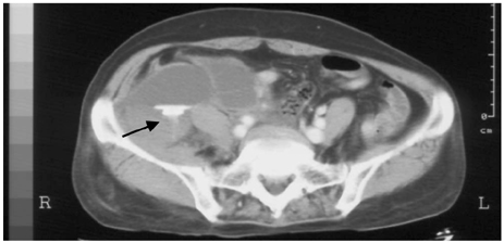
Fig. 1 Initial abdominopevic CT scan. A huge hematoma in the retroperitoneal space was found, and there was contrast enhancement leakage inside of the mass (arrow) implying active bleeding. CT: computed tomography.
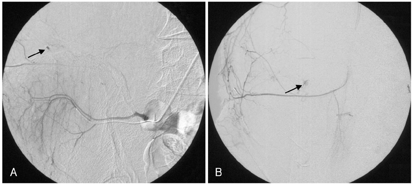
Fig. 2 Initial finding of the angiography. The urgently performed angiography showed two active bleeding foci (arrows): branches of the right circumflex iliac artery (A) and right lumbar artery (B).
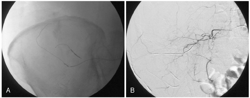
Fig. 3 Coil embolization. A total of 7 microcoils were used to eliminate all bleeding foci, including all collateral arteries. After coil embolization, a final angiography was performed and comfirmed that there was no contrast agent leakage.
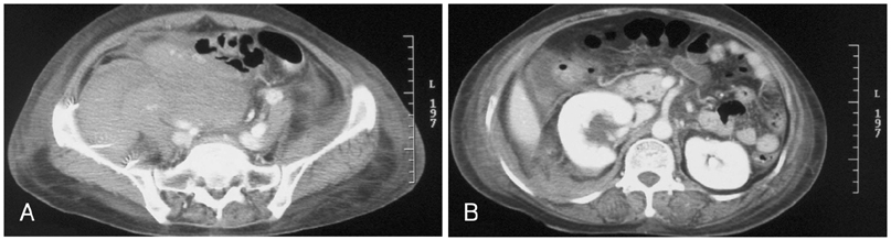
Fig. 4 Follow-up chest CT scan 4 days after the initial embolization. A: the size of the hematoma increased, but no suspicious bleeding sites were found. B: a hydronephrosis in the right kidney developed, probably caused by a right ureteral compression by the mass. CT: computed tomography.
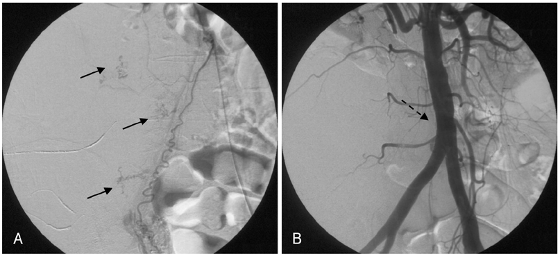
Fig. 5 Another bleeding focus. A: the angiography revealed multiple bleeding foci (dark arrows) from branches of the right ovarian artery. B: after gelfoam injection, angiography showed a totally occluded right ovarian artery at the ostium portion (dotted arrow).
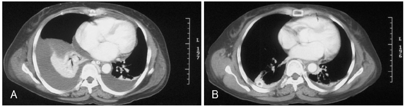
Fig. 6 Both hemothoraces. A: a chest CT scan revealed bilateral hemothoraces, confirmed by diagnostic thoracentesis. A chest tube insertion was performed. B: 9 days later, a follow-up chest CT scan showed a much improved hemothorax. CT: computed tomography.

Fig. 7 One month after the initial chest CT scan. A: the size of the hematoma slightly increased without any bleeding points and the wall of the hematoma was more densely enhanced, indicating that the hematoma had matured. B: the hydroneprosis of the right kidney became more aggravated. CT: computed tomography.
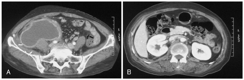
Fig. 8 Three month follow-up chest CT scan. A: the size of the hematoma was decreased. B: the hydronephrosis of the right kidney had nearly resolved. CT: computed tomography.
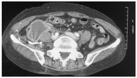
Fig. 9 Seven month follow-up chest CT scan. The hematoma was nearly resolved. CT: computed tomography.
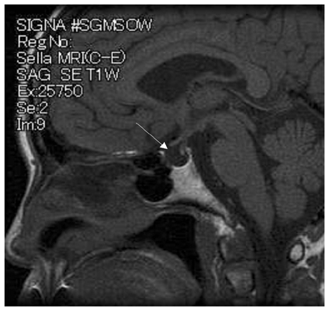
Fig. 10 The sella MRI. A T1-weighted image revealed that the pituitary gland was thin and flat and the sella was empty (arrow). MRI: magnetic resonance imaging.
Reference
-
1. Tappe U, Kristen F, Löffler A, Keller HW. Spontaneous retroperitoneal hematoma in adrenal metastasis. Dtsch Med Wochenschr. 1997. 122:471–474.2. Malik A, Capling R, Bastani B. Enoxaparin-associated retroperitoneal bleeding in two patients with renal insufficiency. Pharmacotherapy. 2005. 25:769–772.3. Ishihara S, Yasuhara H, Ogawa S, Muto T. Successful surgical treatment for spontaneous retroperitoneal hematoma in polycythemia vera: report of a case. Surg Today. 2000. 30:199–201.4. Jeong TK, Jeong GH, Park BS, et al. Dalteparin sodium-associated retroperitoneal hematoma in a patient with diabetic nephropathy. Korean J Med. 2003. 64:322–327.5. Kim HJ, Kim DY, Whang MG, Jo HK. A case of spontaneous retroperitoneal hemorrhage due to iliopsoas muscle hematoma in patient with myocardial infarction receiving intravenous heparin. Korean Circ J. 1998. 28:1798–1801.6. Sreeram S, Lumsden AB, Miller JS, Salam AA, Dodson TF, Smith RB. Retroperitoneal hematoma following femoral arterial catheterization: a serious and often fatal complication. Am Surg. 1993. 59:94–98.7. Chan YC, Morales JP, Reidy JF, Taylor PR. Management of spontaneous and iatrogenic retroperitoneal hemorrhage: conservative management, endovascular intervention or open surgery? Int J Clin Pract. 2007. [Epub ahead of print].8. Baker BH, Baker MS. Indications for exporing the retroperitoneal space. South Med J. 1980. 73:969–970.9. Grimm MR, Vrahas MS, Thromas KA. Pressure-volume characteristics of the intact and disrupted pelvic retroperitoneum. J Trauma. 1998. 44:454–459.10. Kauppila LI. Blood supply of the lower thoracic and lumbosacral regions: postmorterm aortography in 38 young adults. Acta Radiol. 1994. 35:541–544.11. Torres GM, Cernigliaro JG, Abbitt PL, et al. Iliopsoas compartment: normal anatomy and pathologic processes. Radiographics. 1995. 15:1285–1297.12. McCort JJ. Intraperitoneal and retroperitoneal hemorrhage. Radiol Clin North Am. 1976. 14:391–405.13. Berna JD, Zuazu I, Madrigal M, et al. Conservative treatment of large rectus sheath hematoma in patients undergoing anticoagulant therapy. Abdom Imaging. 2000. 25:230–234.14. Dabney A, Bastani B. Enoxaparin-associated severe retroperitoneal bleeding and abdominal compartment syndrome: a report of two cases. Intensive Care Med. 2001. 27:1954–1957.
- Full Text Links
-
- Actions
-
Cited
- CITED
-
- Close
- Share
-
- Similar articles
-
- A Case of Spontaneous Retroperitoneal Hemorrhage due to Iliopsoas Muscle Hematoma in Patient with Myocardial Infarction Receiving Intravenous Heparin
- Transcatheter Arterial Embolization as Treatment for a Life-Threatening Retroperitoneal Hemorrhage Complicating Heparin Therapy
- Spontaneous Retroperitoneal Hemorrhage from Ruptured Suprarenal Artery Aneurysm
- A Case of Spontaneous Hemo-pneumothorax
- A Case of Hemorrhagic Fever with Renal Syndrome Complicated by Retroperitoneal Hematoma and Hemothorax
