Korean J Ophthalmol.
2010 Dec;24(6):377-379. 10.3341/kjo.2010.24.6.377.
Dissociated Horizontal Deviation after Traumatic Brain Injury
- Affiliations
-
- 1Department of Ophthalmology, Korea University College of Medicine, Ansan, Korea. ansaneye@hanmail.net
- 2Department of Neurology, Korea University College of Medicine, Seoul, Korea.
- KMID: 1769095
- DOI: http://doi.org/10.3341/kjo.2010.24.6.377
Abstract
- A 4-year-old boy visited the hospital with exotropia after brain hemorrhage caused by trauma. He had undergone decompressive craniectomy and cranioplasty 18 months prior to presentation at our hospital. An alternate prism cover test showed more than 50 prism diopters (PD) of left exotropia when he was fixing with the right eye and 30 PD of right exotropia when he was fixing with the left eye at near and far distance. On the Hirschberg test, 60 PD of left exotropia was noted in the primary position. Brain computerized tomography imaging performed 18 months prior showed hypodense changes in the right middle cerebral artery and anterior cerebral artery territories. Subfalcian herniation was also noted secondary to swelling of the right hemisphere. The patient underwent a left lateral rectus muscle recession of 7.0 mm and a left medial rectus muscle resection of 3.5 mm. Three weeks after the surgery, the Hirschberg test showed orthotropia. On alternate prism cover testing, 8 PD of left exotropia and 8 PD of right esotropia were noted at distance. We report a patient who developed dissociated horizontal deviation after right subfalcian subdural hemorrhage caused by trauma.
MeSH Terms
Figure
-
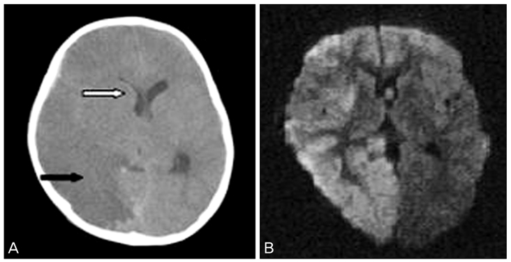
Fig. 1 (A) Computerized tomography showed hypodense change at right middle cerebral artery, both anterior cerebral artery territory (black arrow) and subfalcian herniation due to swelling of right hemisphere (white arrow). (B) Diffuse magnetic resonance imaging showed high signal at the right frontoparietooccipital lobe which means decreased diffusion and acute infarction.
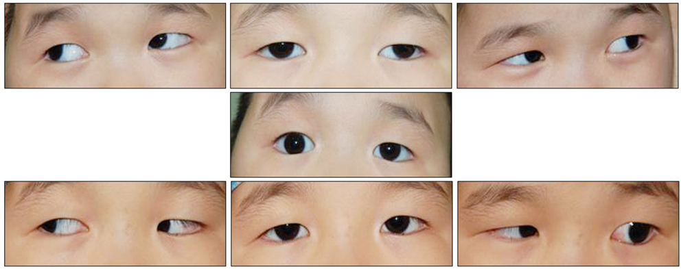
Fig. 2 Note large temporal deviation of left eyeball about 60 prism diopters (PD) fixing with the right eye (top) and right exotropia of 30 PD fixing with the left eye (middle) preoperatively. A small amount of esodeviation in the right eye and 8 PD of exodeviation in the left eye were noted postoperatively (bottom).
Reference
-
1. Brodsky MC, Gräf MH, Kommerell G. The reversed fixation test: a diagnostic test for dissociated horizontal deviation. Arch Ophthalmol. 2005. 123:1083–1087.2. Lee JK, Choi YI, Jin YH. Dissociated horizontal deviation. J Korean Ophthalmol Soc. 1994. 35:480–483.3. Im CS, Lee JB, Kim HS. Dissociated horizontal deviation in the refractive accommodative esotropia with amblyopia. J Korean Ophthalmol Soc. 1997. 38:1268–1272.4. Brodsky MC. Dissociated horizontal deviation: clinical spectrum, pathogenesis, evolutionary underpinnings, diagnosis, treatment, and potential role in the development of infantile esotropia (an American Ophthalmological Society thesis). Trans Am Ophthalmol Soc. 2007. 105:272–293.5. Tychsen L. Causing and curing infantile esotropia in primates: the role of decorrelated binocular input (an American Ophthalmological Society thesis). Trans Am Ophthalmol Soc. 2007. 105:564–593.6. Koh SB, Kim BJ, Lee J, et al. Stereopsis and color vision impairment in patients with right extrastriate cerebral lesions. Eur Neurol. 2008. 60:174–178.
- Full Text Links
-
- Actions
-
Cited
- CITED
-
- Close
- Share
-
- Similar articles
-
- Dissociated Vertical Deviation with Microtia showing Familial Tendency
- The Effect of Unilateral Surgery in Dissociated Vertical Deviation
- Dissociated Horizontal Deviation in the Refractive Accommodative Esotropia with Amblyopia
- Operations on Vertical Rectus Muscles: Their Influence on Horizontal Strabismus
- Asymmetric Bilateral Inferior Oblique Transposition of a Bilateral Inferior Oblique Overaction Associated with Dissociated Vertical Deviation
