A Case of Desmoid Tumor Presenting as Intra-abdominal Abscess
- Affiliations
-
- 1Department of Internal Medicine, Eulji University School of Medicine, Seoul, Korea. sbk1026@eulji.ac.kr
- 2Department of Surgery, Eulji University School of Medicine, Seoul, Korea.
- 3Department of Pathology, Eulji University School of Medicine, Seoul, Korea.
- KMID: 1767693
- DOI: http://doi.org/10.4166/kjg.2009.53.5.315
Abstract
- Desmoid tumor is a rare benign tumor derived from fibrous sheath or musculoaponeurotic structure. The tumor is benign histologically but considered as malignant clinically because it has high propensity on infiltrative growth with local invasion and tendency to recurrence after local excision. Especially, when this tumor happens to be in the intra-abdomen, the prognosis is worse because it can cause intestinal obstruction, ureter obstruction and, fistula formation. It also can invade major vessels in abdomen. This tumor occurs more frequently in patients with familial adenomatous polyposis (FAP), in post-partume women, and at old surgical incision site. However, in this case, the patient had neither previous surgery nor a FAP history. We report a rare case of the young male patient who presented with an acute abdomen and underwent laparotomy and was found to have an intra-abdominal desmoid tumor with abscess formation.
Keyword
MeSH Terms
Figure
-
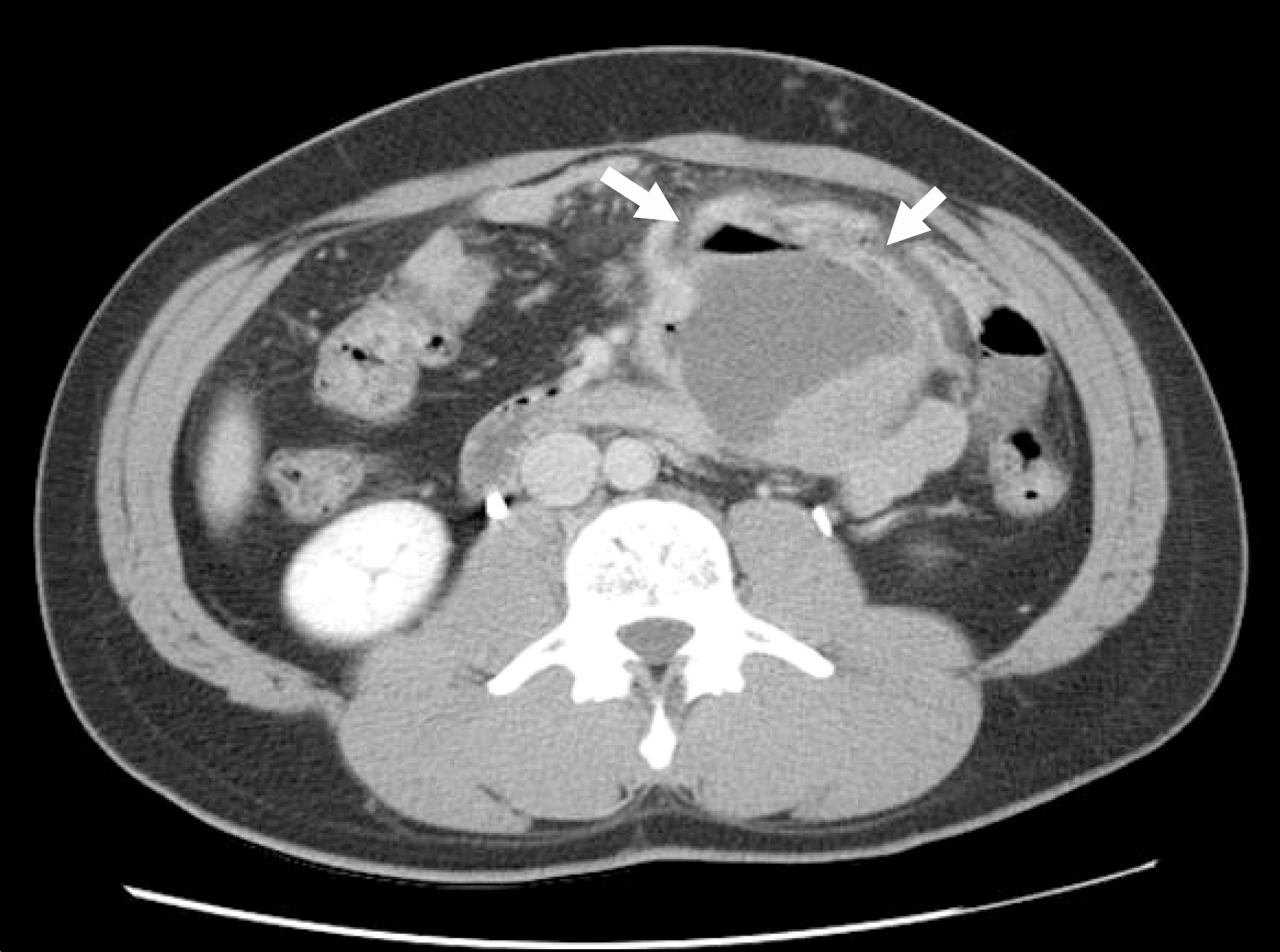
Fig. 1. Abdominal CT finding showed 9×8×10 cm large mass with air-fluid level (arrow) at left lower abdomen.
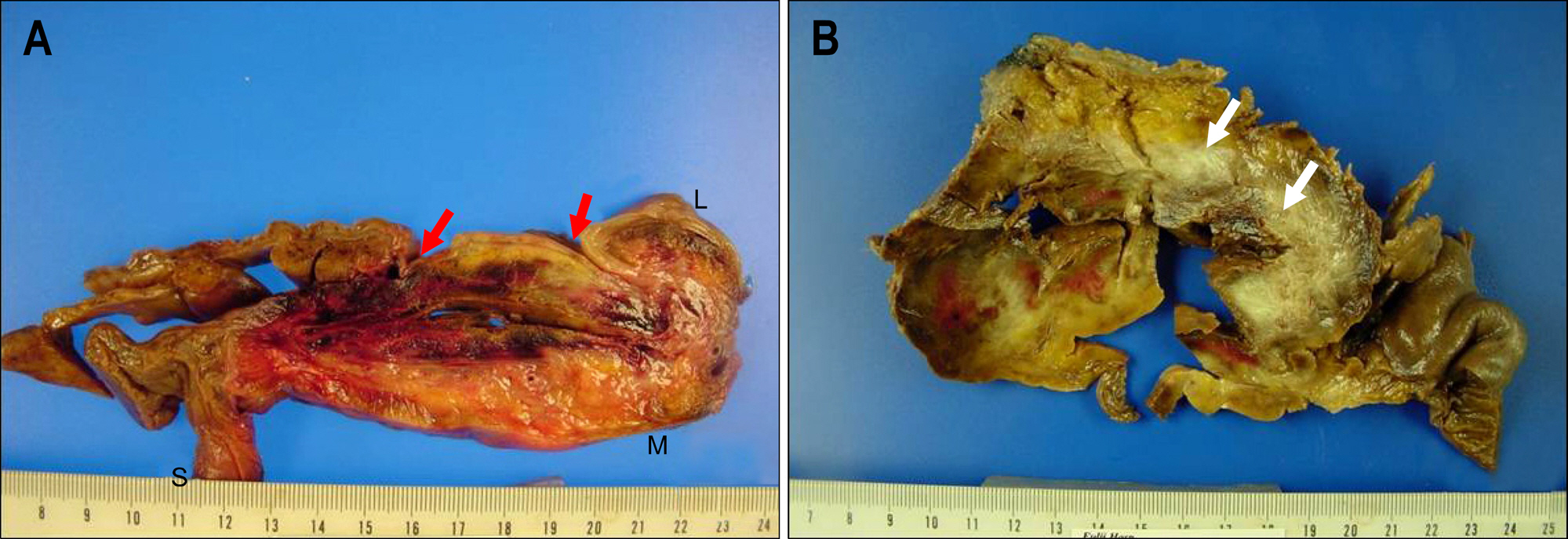
Fig. 2. Gross specimen findings. (A) On opening the large intestine, the mucosa revealed two dimpled foci. Arrows reveal pseudo-diver-ticulum fistular form like traction surrounded by congested firm fibrofatty tissue of mesentery (S : small intestine, L : large intestine, M: mesentery). (B) Arrows show that mesenteric mass between the colon and small intestine with marked congestion and necrotic tissue. It had central irregular space, up to 4 cm in dimension containing congested friable tissue surrounded by dense and firm fibrocollagenous tissue which was continuous to the traction diverticulous foci, and partly surrounded by severe hemorrhagic area.
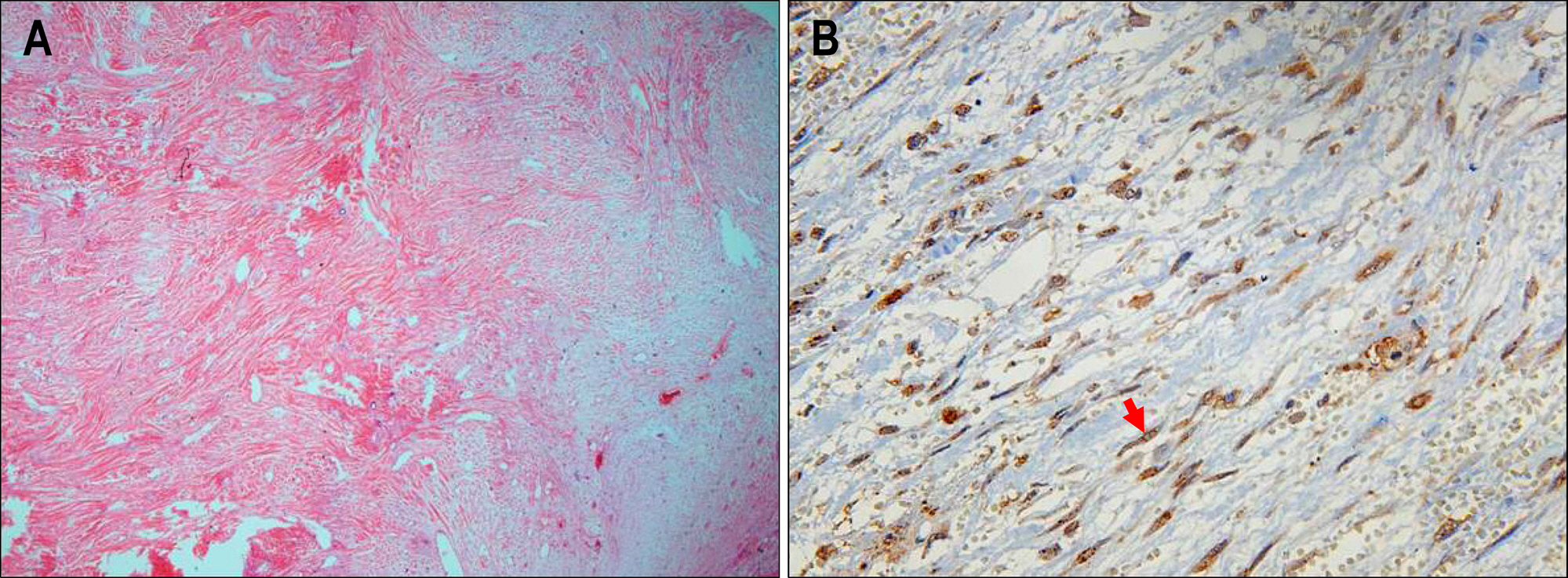
Fig. 3. Microscopic findings. (A) Microscopic finding showed the proliferation of fibroblasts with abundant collagens (H&E stain, ×100). (B) An arrow indicates positive reaction for β-catenin staining in fibroblast (Immunohistochemical stain, ×400).
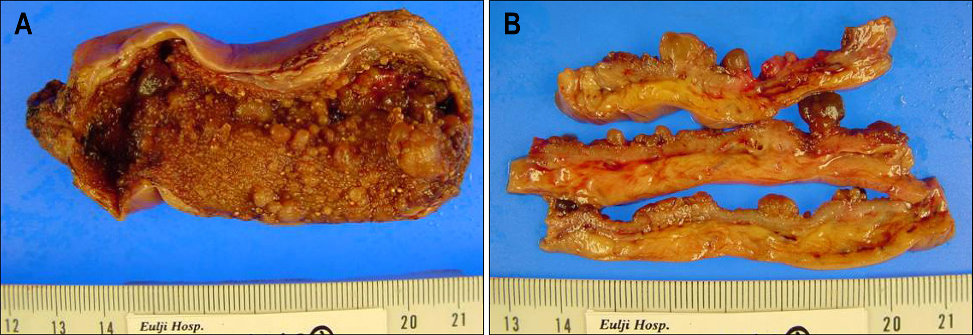
Fig. 4. (A, B) On gross finding, there was no stone but multiple polypoid mucoid lesions in gallbladder.
Reference
-
1. Kulaylat MN, Karakousis CP, Keaney CM, McCorvey D, Bem J, Ambrus Sr JL. Desmoid tumour: a pleomorphic lesion. Eur J Surg Oncol. 1999; 25:487–497.
Article2. Park BH, Kim HJ, Chang YW, et al. Desmoid tumor and duodenal adenoma in a patient with familial adenomatous polyposis: a case report. Korean J Gastrointest Endosc. 2001; 23:32–35.3. Lee HS, Jeon HM, Ok ST, Kim JS, Lee EJ, Kim JS. Unresectable desmoid tumor developing after surgery of F.A.P case report. J Korean Soc Coloproctol. 1998; 14:323–329.4. Cruz-Correa M, Giardiello FM. Familial adenomatous polyposis. Gastrointest Endosc. 2003; 58:885–894.
Article5. Couture J, Mitri A, Lagace R, et al. A germline mutation at the extreme 3' end of the APC gene results in a severe desmoid phenotype and is associated with overexpression of be-ta-catenin in the desmoid tumor. Clin Genet. 2000; 57:205–212.
Article6. Reitamo JJ, Hayry P, Nykyri E, Saxen E. The desmoid tumor. I. Incidence, sex-, age- and anatomical distribution in the Finnish population. Am J Clin Pathol. 1982; 77:665–673.
Article7. Khorsand J, Karakousis CP. Desmoid tumors and their management. Am J Surg. 1985; 149:215–218.
Article8. Eren S. A sporadic abdominal desmoid tumour case presenting with intermittent intestinal obstruction. Eur J Pediatr Surg. 2007; 17:285–288.
Article9. Corbel L, Souissi M, Chretien Y, Dufour B. Desmoid tumor of the mesentery. An uncommon cause of ureteral obstruction. J Radiol. 1992; 73:669–672.10. Smith AJ, Lewis JJ, Merchant NB, Leung DH, Woodruff JM, Brennan MF. Surgical management of intraabdominal desmoid tumours. Br J Surg. 2000; 87:608–613.
Article11. Jalini L, Hemming D, Bhattacharya V. Intraabdominal desmoid tumour presenting with perforation. Surgeon. 2006; 4:114–116.
Article12. Cholongitas E, Koulenti D, Panetsos G, et al. Desmoid tumor presenting as intraabdominal abscess. Dig Dis Sci. 2006; 51:68–69.
Article13. Lopez R, Kemalyan N, Moseley HS, Dennis D, Vetto RM. Problems in diagnosis and management of desmoid tumors. Am J Surg. 1990; 159:450–453.
Article14. Church J, Berk T, Boman BM, et al. Staging intraabdominal desmoid tumors in familial adenomatous polyposis: a search for a uniform approach to a troubling disease. Dis Colon Rectum. 2005; 48:1528–1534.
Article15. Jeong ID, Bang SJ, Shin JW, et al. A case of intraabdominal desmoid tumor after total colectomy in a patient with familial adenomatous polyposis. Korean J Gastrointest Endosc. 2006; 33:50–53.16. Suit H, Spiro I. Radiation in the multidisciplinary management of desmoid tumors. Front Radiat Ther Oncol. 2001; 35:107–119.
Article17. Bus PJ, Verspaget HW, van Krieken JH, et al. Treatment of mesenteric desmoid tumours with the anti-oestrogenic agent toremifene: case histories and an overview of the literature. Eur J Gastroenterol Hepatol. 1999; 11:1179–1183.18. Risum S, Bulow S. Doxorubicin treatment of an intraabdominal desmoid tumour in a patient with familial adenomatous polyposis. Colorectal Dis. 2003; 5:585–586.
Article19. Heidemann J, Ogawa H, Otterson MF, Shidham VB, Binion DG. Antiangiogenic treatment of mesenteric desmoid tumors with toremifene and interferon alfa-2b: report of two cases. Dis Colon Rectum. 2004; 47:118–122.
Article
- Full Text Links
-
- Actions
-
Cited
- CITED
-
- Close
- Share
-
- Similar articles
-
- A Case of Intra-abdominal Desmoid Tumor in a Patient with Familial Adenomatosis Polyposis after Total Colectomy
- Clinical Experience of Partial Resection of Desmoid Tumor and Perforated Small Bowel for Unresectable Desmoid Tumor with Small Bowel Perforation after IPAA for FAP
- A Case of Desmoid Tumor of Rectus Abdominis Muscle Associated with pregnancy
- Desmoid Tumor of the Rectus Abdominis Muscle Associated with Pregnancy
- Intrathoracic Desmoid Tumor Presenting as Multiple Lung Nodules 13 Years after Previous Resection of Abdominal Wall Desmoid Tumor
