Phenotypic Characterization of Ligamentum Flavum Cells from Patients with Ossification of Ligamentum Flavum
- Affiliations
-
- 1Department of Orthopedic and Spinal Surgery, Nanfang Hospital, Southern Medical University, Guangzhou, China. chenjt99@tom.com
- KMID: 1758565
- DOI: http://doi.org/10.3349/ymj.2009.50.3.375
Abstract
-
PURPOSE: The objective of this study was to determine the phenotypic characterization of ligamentum flavum cells from patients with ossification of the ligamentum flavum (OLF).
MATERIALS AND METHODS
Ligamentum flavum tissues were harvested from OLF and non-OLF patients during surgery. OLF and non-OLF cells were isolated from explant cultures. Cultured cells were analyzed using immunofluorescence staining and reverse transcription-polymerase chain reaction.
RESULTS
OLF cells exhibited various appearances compared with the typical fibroblast-like morphology of non-OLF cells. Expressions of collagen type I and collagen type III were observed in OLF and non-OLF cells. OLF cells uniquely expressed osteocalcin, which is a marker for osteoblasts, and collagen type II which is a marker for chondrocytes, whereas they were negative in non-OLF cells.
CONCLUSION
These findings indicate that OLF cells have phenotypic characterization of osteoblasts and chondrocytes which could play a role in the pathophysiology of OLF.
Keyword
MeSH Terms
-
Adolescent
Adult
Aged
Cells, Cultured
Collagen Type I/genetics/metabolism
Collagen Type II/genetics/metabolism
Collagen Type VI/genetics/metabolism
Female
Humans
Ligamentum Flavum/metabolism/*pathology
Male
Microscopy, Fluorescence
Middle Aged
Ossification, Heterotopic/metabolism/*pathology
Osteocalcin/genetics/metabolism
Reverse Transcriptase Polymerase Chain Reaction
Young Adult
Figure
-
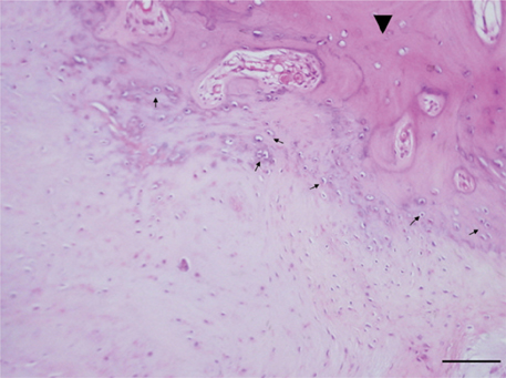
Fig. 1 Histological characterization of the OLF. Ossification (arrowhead) and chondroid cells (arrow) were observed in the ossified ligamentum flavum (hematoxylin and eosin stain, bar = 100 µm).
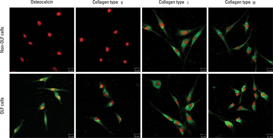
Fig. 2 Immunofluorescence analysis of OLF cells and non-OLF cells. Immunofluorescence was visualized using FITC (green) and the nuclei by PI (red). OLF cells expressed osteocalcin, collagen type I, II and III; non-OLF cells expressed collagen type I and III, but not osteocalcin and collagen type II (bar = 20 µm).
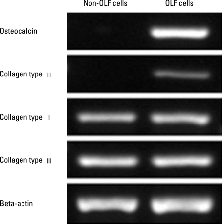
Fig. 3 Gene expression analysis for osteocalcin, collagen type I, collagen type II and collagen type III in OLF cells and non-OLF cells by RT-PCR. Data shown are agarose gel electrophoresis of amplification products.
Cited by 1 articles
-
Inter- and Intra-Observer Variability of the Volume of Cervical Ossification of the Posterior Longitudinal Ligament Using Medical Image Processing Software
Dong Ah Shin, Gyu Yeul Ji, Chang Hyun Oh, Keung Nyun Kim, Do Heum Yoon, Hyunchul Shin
J Korean Neurosurg Soc. 2017;60(4):441-447. doi: 10.3340/jkns.2015.0708.014.
Reference
-
1. al-Orainy IA, Kolawole T. Ossification of the ligament flavum. Eur J Radiol. 1998. 29:76–82.
Article2. Kuh SU, Kim YS, Cho YE, Jin BH, Kim KS, Yoon YS, et al. Contributing factors affecting the prognosis surgical outcome for thoracic OLF. Eur Spine J. 2006. 15:485–491.
Article3. Pascal-Moussellard H, Cabre P, Smadja D, Catonné Y. Symptomatic ossification of the ligamentum flavum: a clinical series from the French Antilles. Spine. 2005. 30:E400–E405.4. Aizawa T, Sato T, Sasaki H, Kusakabe T, Morozumi N, Kokubun S. Thoracic myelopathy caused by ossification of the ligamentum flavum: clinical features and surgical results in the Japanese population. J Neurosurg Spine. 2006. 5:514–519.
Article5. Tokala DP, Lam KS, Prince HG. Ossification of the proximal thoracic ligamenta flava causing acute myelopathy in a Caucasian: case report and literature review. Spinal Cord. 2007. 45:310–313.
Article6. Nishiura I, Isozumi T, Nishihara K, Handa H, Koyama T. Surgical approach to ossification of the thoracic yellow ligament. Surg Neurol. 1999. 51:368–372.
Article7. Shiigi E, Sugiyama T, Tanaka H, Murata H, Shirakura Y, Kawai S. Possible involvement of vitamin D receptor gene polymorphism in male patients with ossification of spinal ligaments. J Bone Miner Metab. 2001. 19:308–311.
Article8. Kong Q, Ma X, Li F, Guo Z, Qi Q, Li W, et al. COL6A1 polymorphisms associated with ossification of the ligamentum flavum and ossification of the posterior longitudinal ligament. Spine. 2007. 32:2834–2838.
Article9. Tsukamoto N, Maeda T, Miura H, Jingushi S, Hosokawa A, Harimaya K, et al. Repetitive tensile stress to rat caudal vertebrae inducing cartilage formation in the spinal ligaments: a possible role of mechanical stress in the development of ossification of the spinal ligaments. J Neurosurg Spine. 2006. 5:234–242.
Article10. Maigne JY, Ayral X, Guérin-Surville H. Frequency and size of ossifications in the caudal attachments of the ligamentum flavum of the thoracic spine. Role of rotatory strains in their development. An anatomic study of 121 spines. Surg Radiol Anat. 1992. 14:119–124.
Article11. Shirakura Y, Sugiyama T, Tanaka H, Taguchi T, Kawai S. Hyperleptinemia in female patients with ossification of spinal ligaments. Biochem Biophys Res Commun. 2000. 267:752–755.
Article12. Terakado A, Tagawa M, Goto S, Yamazaki M, Moriya H, Fujimura S. Elevation of alkaline phosphatase activity induced by parathyroid hormone in osteoblast-like cells from the spinal hyperostotic mouse TWY (twy/twy). Calcif Tissue Int. 1995. 56:135–139.
Article13. Hayashi K, Ishidou Y, Yonemori K, Nagamine T, Origuchi N, Maeda S, et al. Expression and localization of bone morphogenetic proteins (BMPs) and BMP receptors in ossification of the ligamentum flavum. Bone. 1997. 21:23–30.
Article14. Sakamoto R, Ikata T, Murase M, Hasegawa T, Fukushima T, Hizawa K. Comparative study between magnetic resonance imaging and histopathologic findings in ossification or calcification of ligaments. Spine. 1991. 16:1253–1261.
Article15. Tanaka H, Nagai E, Murata H, Tsubone T, Shirakura Y, Sugiyama T, et al. Involvement of bone morphogenic protein-2 (BMP-2) in the pathological ossification process of the spinal ligament. Rheumatology (Oxford). 2001. 40:1163–1168.
Article16. Yayama T, Uchida K, Kobayashi S, Kokubo Y, Sato R, Nakajima H, et al. Thoracic ossification of the human ligamentum flavum: histopathological and immunohistochemical findings around the ossified lesion. J Neurosurg Spine. 2007. 7:184–193.
Article17. Okada K, Oka S, Tohge K, Ono K, Yonenobu K, Hosoya T. Thoracic myelopathy caused by ossification of the ligamentum flavum. Clinicopathologic study and surgical treatment. Spine. 1991. 16:280–287.
Article18. Specchia N, Pagnotta A, Gigante A, Logroscino G, Toesca A. Characterization of cultured human ligamentum flavum cells in lumbar spine stenosis. J Orthop Res. 2001. 19:294–300.
Article19. zur Nieden NI, Kempka G, Ahr HJ. In vitro differentiation of embryonic stem cells into mineralized osteoblasts. Differentiation. 2003. 71:18–27.
Article20. Huh YH, Ryu JH, Chun JS. Regulation of type II collagen expression by histone deacetylase in articular chondrocytes. J Biol Chem. 2007. 282:17123–17131.
Article21. Pantazis G, Tsitsopoulos P, Bibis A, Mihas C, Chatzistamou I, Kouzelis C. Symptomatic ossification of the ligamentum flavum at the lumbar spine: a retrospective study. Spine. 2008. 33:306–311.22. Inamasu J, Guiot BH, Sachs DC. Ossification of the posterior longitudinal ligament: an update on its biology, epidemiology, and natural history. Neurosurgery. 2006. 58:1027–1039.
Article23. Vanden Bossche L, Vanderstraeten G. Heterotopic ossification: a review. J Rehabil Med. 2005. 37:129–136.24. Nakatani T, Marui T, Hitora T, Doita M, Nishida K, Kurosaka M. Mechanical stretching force promotes collagen synthesis by cultured cells from human ligamentum flavum via transforming growth factor-beta1. J Orthop Res. 2002. 20:1380–1386.
Article
- Full Text Links
-
- Actions
-
Cited
- CITED
-
- Close
- Share
-
- Similar articles
-
- Thoracic Myelopathy Due to Ossification of Ligamentum Flavum: Three cases Report
- As a Cause of Myelopathy in the Lower Thracic Spines ): Two Cases Report
- Ossified Ligamentum Flavum causing Cervical Myelopathy
- Myelopathy due to Ossification of Ligamentum Flavum
- Sacral Radiculopathy Due to Ossification of Ligamentum Flavum and Posterior Longitudinal Ligament: One Case Report
