J Cerebrovasc Endovasc Neurosurg.
2014 Dec;16(4):358-363. 10.7461/jcen.2014.16.4.358.
Volume Measurement of the Intracranial Aneurysm: A Discussion and Comparison of the Alternatives to Manual Segmentation
- Affiliations
-
- 1Department of Radiology, Queen Mary Hospital, Hong Kong S.A.R, China. victorchansh@gmail.com
- 2Department of Neurosurgery, Kwong Wah Hospital, Hong Kong S.A.R, China.
- KMID: 1738341
- DOI: http://doi.org/10.7461/jcen.2014.16.4.358
Abstract
OBJECTIVE
Several modalities are available for volumetric measurement of the intracranial aneurysm. We discuss the challenges involved in manual segmentation, and analyze the application of alternative methods using automatic segmentation and geometric formulae in measurement of aneurysm volumes and coil packing density.
METHODS
The volumes and morphology of 38 aneurysms treated with endovascular coiling at a single center were measured using three-dimensional rotational angiography (3DRA) reconstruction software using automatic segmentation. Aneurysm volumes were also calculated from their height, width, depth, size of neck, and assumed shape in 3DRA images using simple geometric formulae. The aneurysm volumes were dichotomized as "small" or "large" using the median volume of the studied population (54 mm3) measured by automatic segmentation as the cut-off value for further statistical analysis.
RESULTS
A greater proportion of aneurysms were categorized as being "small" when geometric formulae were applied. The median aneurysm volumes obtained were 54.5 mm3 by 3DRA software, and 30.6 mm3 using mathematical equations. An underestimation of aneurysm volume with a resultant overestimation in the calculated coil packing density (p = 0.002) was observed.
CONCLUSION
Caution must be exercised in the application of simple geometric formulae in the management of intracranial aneurysms as volumes may potentially be underestimated and packing densities falsely elevated. Future research should focus on validation of automatic segmentation in volumetric measurement and improving its accuracy to enhance its application in clinical practice.
Keyword
Figure
-
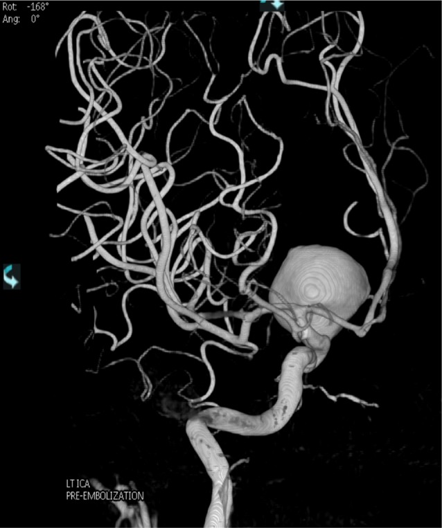
Fig. 1 A giant unruptured left internal carotid artery aneurysm prior to embolization.
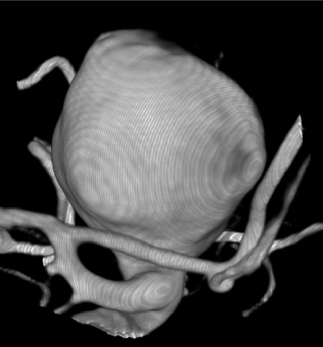
Fig. 2 The aneurysm is identified and isolated with its parent vessels.
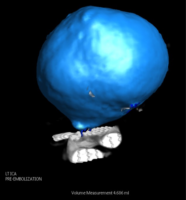
Fig. 3 The volume is measured by automatic segmentation via reconstruction software.
Cited by 1 articles
-
An Efficient Method for Aneurysm Volume Quantification Applicable in Any Shape and Modalities
Jaewoo Chung, Jung Ho Ko
J Korean Neurosurg Soc. 2021;64(4):514-523. doi: 10.3340/jkns.2020.0255.
Reference
-
1. Bescós JO, Slob MJ, Slump CH, Sluzewski M, van Rooij WJ. Volume measurement of intracranial aneurysms from 3D rotational angiography: improvement of accuracy by gradient edge detection. AJNR Am J Neuroradiol. 2005; Nov-Dec. 26(10):2569–2572. PMID: 16286402.2. Brilstra EH, Rinkel GJ, van der Graaf Y, van Rooij WJ, Algra A. Treatment of intracranial aneurysms by embolization with coils: a systematic review. Stroke. 1999; 2. 30(2):470–476. PMID: 9933290.3. Bogunović H, Pozo JM, Villa-Uriol MC, Majoie CB, van den Berg R, Gratama van Andel HA, et al. Automated segmentation of cerebral vasculature with aneurysms in 3DRA and TOF-MRA using geodesic active regions: an evaluation study. Med Phys. 2011; 1. 38(1):210–222. PMID: 21361189.
Article4. Molyneux A, Kerr R, Stratton I, Sandercock P, Clarke M, Shrimpton J, et al. International Subarachnoid aneurysm Trial (ISAT) of neurosurgical clipping versus endovascular coiling in 2143 patients with ruptured intracranial aneurysms: a randomised trial. Lancet. 2002; 10. 360(9342):1267–1274. PMID: 12414200.
Article5. Piotin M, Spelle L, Mounayer C, Salles-Rezende MT, Giansante-Abud D, Vanzin-Santos R, et al. Intracranial aneurysms: treatment with bare platinum coils - aneurysm packing, complex coils and angiographic recurrence. Radiology. 2007; 5. 243(2):500–508. PMID: 17293572.6. Ryu CW, Kwon OK, Koh JS, Kim EJ. Analysis of aneurysm rupture in relation to the geometric indices: aspect ratio, volume and volume-to-neck ratio. Neuroradiology. 2011; 11. 53(11):883–889. PMID: 21107548.
Article7. Sadato A, Hayakawa M, Tanaka T, Hirose Y. Comparison of cerebral aneurysm volumes as determined by digitally measured 3D rotational angiography and approximation from three diameters. Interv Neuroradiol. 2011; 6. 17(2):154–158. PMID: 21696652.
Article8. Sluzewski M, van Rooij WJ, Slob MJ, Bescos JO, Slump CH, Wijnalda D. Relation between aneurysm volume, packing, and compaction in 145 cerebral aneurysms treated with coils. Radiology. 2004; 6. 231(3):653–658. PMID: 15118115.
Article9. van Rooij WJ, Sluzewski M. Opinion: imaging follow-up after coiling of intracranial aneurysms. AJNR Am J Neuroradiol. 2009; 10. 30(9):1646–1648. PMID: 19617448.
- Full Text Links
-
- Actions
-
Cited
- CITED
-
- Close
- Share
-
- Similar articles
-
- Inter-observer and intra-observer reliability between manual segmentation and semi-automated segmentation for carotid vessel wall volume measurements on three-dimensional ultrasonography
- Intracranial "De Novo" Aneurysms: Case Report
- Measurement of Apparent Diffusion Coefficient Values from Diffusion-Weighted MRI: A Comparison of Manual and Semiautomatic Segmentation Methods
- Stent-Assisted Coil Trapping in a Manual Internal Carotid Artery Compression Test for the Treatment of a Fusiform Dissecting Aneurysm
- Diagnostic Availability of Optical Coherence Angiography in Type 1 and 2 Choroidal Neovascularization
