Korean J Lab Med.
2011 Jul;31(3):201-204. 10.3343/kjlm.2011.31.3.201.
Subcutaneous Phaeohyphomycosis Caused by Phaeoacremonium Species in a Kidney Transplant Patient: The First Case in Korea
- Affiliations
-
- 1Department of Laboratory Medicine and Research Institute of Bacterial Resistance, Yonsei University College of Medicine, Seoul, Korea. deyong@yuhs.ac
- 2Department of General Surgery, Yonsei University College of Medicine, Seoul, Korea.
- 3Department of Pathology, Yonsei University College of Medicine, Seoul, Korea.
- KMID: 1735855
- DOI: http://doi.org/10.3343/kjlm.2011.31.3.201
Abstract
- Phaeohyphomycosis is a subcutaneous infection caused by dark pigmented fungi, including fungi of the species Phaeoacremonium, Alternaria, Exophiala, and Pyrenochaeta. In August 2005, a 54-yr-old man who had received a renal transplant 5 yr ago was admitted to our hospital with a subcutaneous mass on the third finger of the right hand; the mass had been present for several months. He had been receiving immunosuppressive agents for several years. He underwent excision of the mass, which was followed by aspiration of the wound for bacterial and fungal cultures. Many fungal hyphae were observed on the histology slide treated with periodic acid-Schiff stain. A few white waxy colonies with a woolly texture grew on the Sabouraud dextrose agar at 30degrees C and changed to dark brown in color. Nucleotide sequencing of internal transcribed spacer regions revealed 100% homology to the Phaeoacremonium aleophilum anamorph and Togninia minima teleomorph (514 bp/514 bp). The patient completely recovered after wide surgical excision. Here, we report the first case of phaeohyphomycosis caused by Phaeoacremonium species in a kidney transplant patient in Korea.
Keyword
MeSH Terms
Figure
-
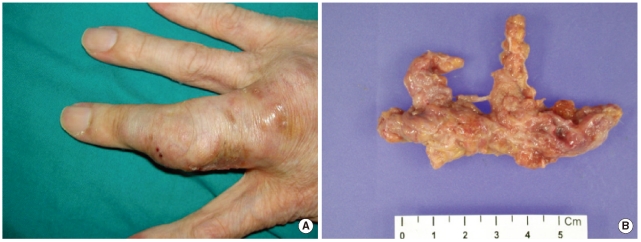
Fig. 1 (A) Subcutaneous lesion on the third finger of the right hand; (B) 6.5×3×1.2 cm-sized mass obtained after removal of the lesion.
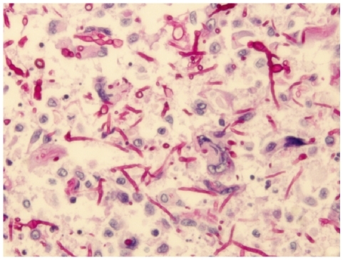
Fig. 2 Periodic acid-Schiff stain showed many fungal hyphae under the microscope (×400).
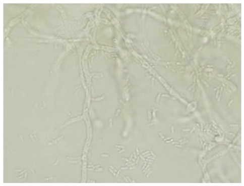
Fig. 3 On the slide culture of the specimen, many fungal hyphae were septate, and conidiophores were observed to be short and usually unbranched (×400).
Reference
-
1. Crous PW, Gams W, Wingfield MJ, Van Wyk PS. Phaeoacremonium gen. nov. associated with wilt and decline diseases of woody hosts and human infections. Mycologia. 1996; 88:786–796.2. Aroca A, Raposo R, Lunello P. A biomarker for the identification of four Phaeoacremonium species using the beta-tubulin gene as the target sequence. Appl Microbiol Biotechnol. 2008; 80:1131–1140. PMID: 18719899.3. Aguilar-Donis A, Torres-Guerrero E, Arenas-Guzman R, Hernandez-Hernandez F, Lopez-Garcia L, Criales-Vera S, et al. Mycetoma caused by Phaeoacremonium parasiticum- a case confirmed with B-tubulin sequence analysis. Mycoses Epub 2010 Aug 4. http://www.ncbi.nlm.nih.gov/pubmed/20701685.4. Essakhi S, Mugnai L, Crous PW, Groenewald JZ, Surico G. Molecular and phenotypic characterisation of novel Phaeoacremonium species isolated from esca diseased grapevines. Persoonia. 2008; 21:119–134. PMID: 20396582.5. Mostert L, Groenewald JZ, Summerbell RC, Robert V, Sutton DA, Padhye AA, et al. Species of Phaeoacremonium associated with infections in humans and environmental reservoirs in infected woody plants. J Clin Microbiol. 2005; 43:1752–1767. PMID: 15814996.6. Naggie S, Perfect JR. Molds: hyalohyphomycosis, phaeohyphomycosis, and zygomycosis. Clin Chest Med. 2009; 30:337–353. vii–viii. PMID: 19375639.
Article7. Guarro J, Alves SH, Gene J, Grazziotin NA, Mazzuco R, Dalmagro C, et al. Two cases of subcutaneous infection due to Phaeoacremonium spp. J Clin Microbiol. 2003; 41:1332–1336. PMID: 12624080.8. Baradkar VP, Mathur M, Kumar S. Phaeohyphomycosis of subcutaneous tissue caused by Phaeoacremonium parasiticum. Indian J Med Microbiol. 2009; 27:66–69. PMID: 19172066.9. Kumar KK, Hallikeri K. Phaeohyphomycosis. Indian J Pathol Microbiol. 2008; 51:556–558. PMID: 19008596.
Article10. Clinical and Laboratory Standerds Institute. Interpretive criteria for identification of bacteria and fungi by DNA target sequencing (MM18-A) 2008.11. Damm U, Mostert L, Crous PW, Fourie PH. Novel Phaeoacremonium species associated with necrotic wood of Prunus trees. Persoonia. 2008; 20:87–102. PMID: 20467488.12. Farina C, Gotti E, Mouniee D, Boiron P, Goglio A. Phaeoacremonium parasiticum subcutaneous infection in a kidney-transplanted patient successfully treated by surgery. Transpl Infect Dis. 2007; 9:253–255. PMID: 17605749.13. Reblova M, Seifert KA. A new fungal genus, Teracosphaeria, with a phialophora-like anamorph (Sordariomycetes, Ascomycota). Mycol Res. 2007; 111:287–298. PMID: 17363235.14. Das S, Saha R, Dar SA, Ramachandran VG. Acremonium species: a review of the etiological agents of emerging hyalohyphomycosis. Mycopathologia. 2010; 170:361–375. PMID: 20577905.15. de Hoog GS, Mayser P, Haase G, Horre R, Horrevorts AM. A new species, Phialophora europaea, causing superficial infections in humans. Mycoses. 2000; 43:409–416. PMID: 11204358.16. Larsen CG, Arendrup MC, Krarup E, Pedersen M, Thybo S, Larsen FG. Subcutaneous phaeohyphomycosis in a renal transplant recipient successfully treated with voriconazole. Acta Derm Venereol. 2009; 89:657–658. PMID: 19997708.
- Full Text Links
-
- Actions
-
Cited
- CITED
-
- Close
- Share
-
- Similar articles
-
- A Case of Subcutaneous Phaeohyphomycosis Caused by Exophiala oligosperma Showing Multiple Cysts
- Cutaneous phaeohyphomycosis in renal transplant recipient
- A Case of Cutaneous Phaeohyphomycosis in an Immunocompetent Patient Caused by Curvularia Species: Case Report and Review of the Literature
- A Case of Subcutaneous and Intranasal Phaeohyphomycosis Caused by Microsphaeropsis arundinis in an Immunocompromised Patient Misdiagnosed with Mucormycosis
- Phaeohyphomycosis Due to Exophiala dermatitidis Successfully Treated with Itraconazole
