Yonsei Med J.
2005 Jun;46(3):372-378. 10.3349/ymj.2005.46.3.372.
Cortical Margining Capabilities of Fins Associated with Ventral Cervical Spine Instrumentation
- Affiliations
-
- 1Department of Neurosurgery, Yonsei University College of Medicine, Seoul, Korea. bhjin61@yumc.yonsei.ac.kr
- KMID: 1734072
- DOI: http://doi.org/10.3349/ymj.2005.46.3.372
Abstract
- Fins incorporated into the design of a dynamic cervical spine implant have been employed to enhance axial load- bearing ability, yet their true biomechanical advantages, if any, have not been defined. Therefore, the goal of this study was to assess the biomechanical and axial load-bearing contributions of the fin components of the DOC ventral cervical stabilization system. Eighteen fresh cadaveric thoracic vertebrae (T1-T3) were obtained. Three test conditions were devised and studied: Condition A (DOC implants with fins were placed against the superior endplate and bone screws were not inserted) ; Condition B (DOC implant without fins was placed and bone screws were inserted) ; and Condition C (DOC implant with fins were placed against the superior endplate and bone screws were inserted). Specimens were tested by applying a pure axial compressive load to the superior platform of the DOC construct, and load-displacement data were collected. Condition C specimens had the greatest stiffness (459 +/- 80N/mm) and yield load (526 +/- 168N). Condition A specimens were the least stiff (266 +/- 53N/mm), and had the smallest yield loads (180 +/- 54N). The yield load of condition A plus condition B was approximately equal to that of condition C, with condition A contributing about one-third and condition B contributing two-thirds of the overall load-bearing capacity. Although the screws alone contributed to a substantial portion of axial load-bearing ability, the addition of the fins further increased load-bearing capabilities.
Keyword
MeSH Terms
Figure
-
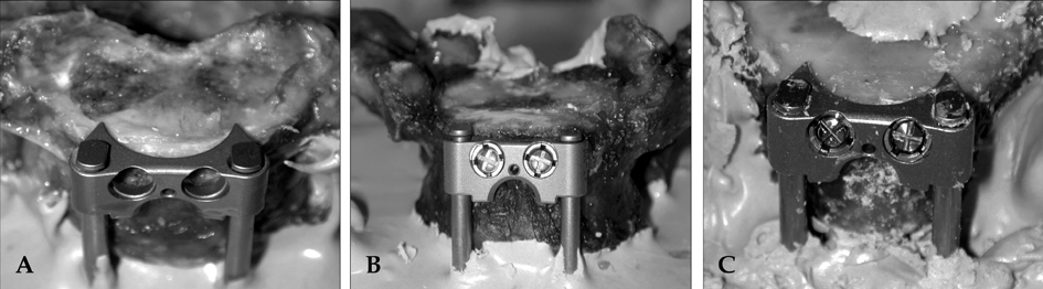
Fig. 1 A: Condition A instrumentation; platform was placed so that the fins were resting against the superior endplate, and no screws were inserted. B: Condition B instrumentation; platforms without fins were placed on the vertebral bodies, and two screws were inserted. C: Condition C instrumentation; platform was placed so that the fins were resting against the superior endplate, and two screws were inserted.

Fig. 2 Specimen setup on materials testing system; a probe was used to axially compress the superior DOC platform.
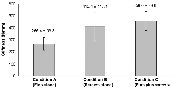
Fig. 3 Bar graph showing the stiffness values of the three instrumented conditions.
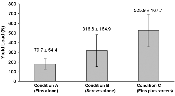
Fig. 4 Bar graph showing the yield loads of the three instrumented conditions.
Reference
-
1. DiAngelo DJ, Foley KT, Vossel KA, Rampersaud YR, Jansen TH. Anterior cervical plating reverses load transfer through multilevel strut-grafts. Spine. 2000. 25:783–795.2. Wang JL, Panjabi MM, Isomi T. The role of bone graft force in stabilizing the multilevel anterior cervical spine plate system. Spine. 2000. 25:1649–1654.3. Brodke DS, Gollogly S, Mohr RA, Nguyen BH, Dailey AT, Bachus KN. Dynamic cervical plates: Biomechanical evaluation of load sharing and stiffness. Spine. 2001. 26:1324–1329.4. Clausen JD, Ryken TC, Traynelis VC, Sawin PD, Dexter F, Goel VJ. Biomechanical evaluation of Caspar and cervical spine locking plate systems in a cadaveric model. J Neurosurg. 1996. 84:1039–1045.5. Grubb MR, Currier BL, Shin JS, Bonin V, Grabowski JJ, Chao EYS. Biomechanical evaluation of anterior cervical spine stabilization. Spine. 1998. 23:886–892.6. Kanayama M, Cunningham MS, Weis JC, Parker LM, Kaneda K, McAfee PC. The effect of rigid spinal instrumentation and solid bony fusion on spinal kinematics. Spine. 1998. 23:767–773.7. Koh YD, Lim TH, You JW, Eck J, An HS. A biomechanical comparison of modern anterior and posterior fixation of the cervical spine. Spine. 2001. 26:15–21.8. Spivak JM, Chen D, Kummer FJ. The effect of locking fixation screws on the stability of anterior cervical plating. Spine. 1999. 24:334–338.9. Brown JA, Havel P, Ebrahein N, Greenblatt SH, Jackson WT. Cervical stabilization by plate and bone fusion. Spine. 1988. 13:236–240.10. Newman M. The outcome of pseudoarthrosis after cervical anterior fusion. Spine. 1993. 18:2380–2382.11. Vaccaro AR, Falatyn SP, Scuderi GJ. Early failure of long segment anterior cervical plate fixation. J Spinal Disord. 1998. 11:410–415.12. Benzel EC. Biomechanics of spine stabilization. 2001. American association of neurological surgeons;155–170. 431–451.13. Wang JC, Zou D, Yuan H, Yoo J. A biomechanical evaluation of graft loading characteristics for anterior cervical discectomy and fusion. Spine. 1998. 23:2450–2454.14. Robinson RA, Walker AE, Ferlic DC, Wiecking DK. The results of anterior interbody fusion of the cervical spine. J Bone Joint Surg. 1962. 44:1569–1586.15. Silva MJ, Keaveny TM, Hayes WC. Load sharing between the shell and centrum in the lumbar vertebral body. Spine. 1997. 22:140–150.16. Brinckmann P, Frobin W, Hierholzer E, Horst M. Deformation of the vertebral endplate under axial loading of the spine. Spine. 1983. 8:851–856.17. Mazess RB. Fracture risk. A role for compact bone. Calc Tiss Int. 1990. 47:191–193.18. Mosekilde L, Mosekilde L. Normal vertebral body size and compressive strength: Relations to age and to vertebral and iliac trabecular bone compressive strength. Bone. 1986. 7:207–212.19. Mosekilde L, Viidik A, Mosekilde L. Correlation between the compressive strength of iliac and vertebral trabecular bone in normal individuals. Bone. 1985. 6:291–295.20. Edwards WT, Zheng Y, Ferrara LA, Yuan HA. Structural features and thickness of the vertebral cortex in the thoracolumbar spine. Spine. 2001. 26:218–225.21. Horst M, Brinckmann P. Measurement of the distribution of axial stress on the end-plate of the vertebral body. Spine. 1981. 6:217–232.22. Silva MJ, Wang C, Keaveny TM, Hayes WC. Direct and computed tomography thickness measurements of the human, lumbar vertebral shell and endplate. Bone. 1994. 15:409–414.23. Burr DB, Yang KH, Haley M, Wang HC. Takashi H, editor. Morphological changes and stress redistribution in osteoporotic spine. Spinal Disorders in Aging. 1994. Tokyo: Springer-Verlag.24. Rockoff SD, Sweet E, Bleustein J. The relative contribution of trabecular and cortical bone to the strength of human lumbar vertebrae. Calcif Tissue Res. 1969. 3:163–175.25. Yoganadan N, Myklebust JB, Cusick JF, Wilson CR, Sances A. Functional biomechanics of the thoracolumbar vertebral cortex. Clin Biomech. 1988. 3:11–18.
- Full Text Links
-
- Actions
-
Cited
- CITED
-
- Close
- Share
-
- Similar articles
-
- Retraction: Cortical Margining Capabilities of Fins Associated with Ventral Cervical Spine Instrumentation
- Anterior Cervical Fusion with the PCB Instrumentation (Cervical Plate Cage System) in Degenerative Cervical Disease
- Delayed Cervical Epidural Abscess after Instrumentation
- Bilateral Pedicle and Crossed Translaminar Screws in C2
- Complications of Anterior and Posterior Cervical Spine Surgery
